Bilder
Videos
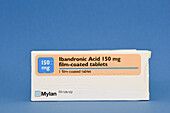
13674429 - Ibandronic acid osteoporosis drug

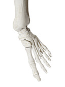
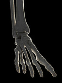
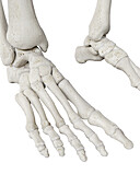
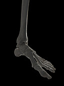
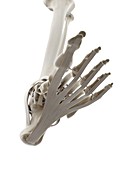
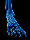
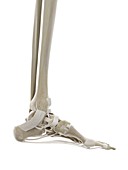
13600710 - Foot bones, illustration
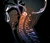
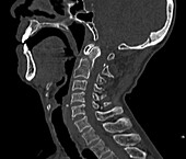
13600164 - Odontoid process fracture, CT scan

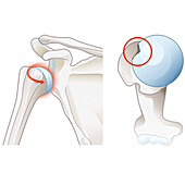
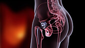
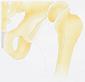
13585898 - Osteoarthritis of the hip, illustration
14078633 - Subacromial bursitis of the shoulder, illustration
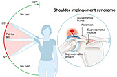
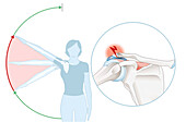
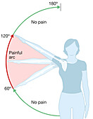
14078614 - Shoulder impingement, illustration
14078612 - Shoulder impingement, illustration
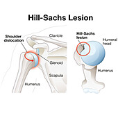
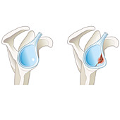
14078575 - Hill-Sachs lesion of the shoulder, illustration
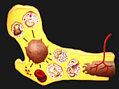
13632784 - Blood cell production, illustration
13585376 - Gout
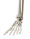
13600802 - Foot bones, illustration
13600800 - Foot bones, illustration
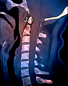
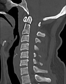
13600168 - Odontoid process fracture, CT scan
13619671 - Knee meniscus injury, illustration
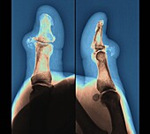
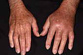
13453242 - Pseudogout of a thumb, X-ray scans
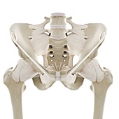
13224524 - Ligaments of the hip, illustration
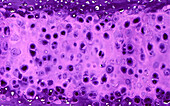
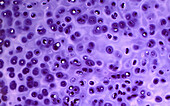
13732524 - Cartilage cells, light micrograph
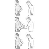
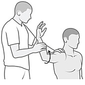
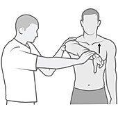
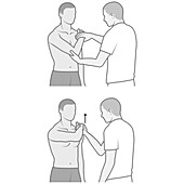
14078608 - Shoulder examination, illustration
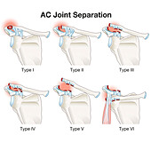
14078544 - AC joint separation, illustration
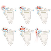
14078543 - AC joint separation, illustration
13674441 - Ibandronic acid osteoporosis drug
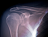
13600398 - Calcific tendinitis, X-ray
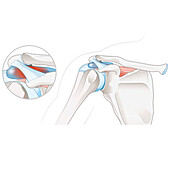
14078635 - Subacromial bursitis of the shoulder, illustration
14078607 - Shoulder examination, illustration
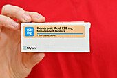
13674443 - Ibandronic acid osteoporosis drug
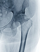
13600839 - Osteoarthritis of the hip, X-ray
14078610 - Shoulder examination, illustration
14078557 - Bony bankart lesion in the shoulder, illustration
13600711 - Foot bones, illustration

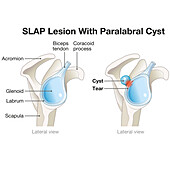
14078628 - SLAP lesion paralabral cyst in the shoulder, illustration
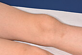
13954611 - Swollen right knee caused by ligament strain
13600803 - Foot bones, illustration
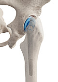
13585903 - Prosthetic hip joint, illustration
14078613 - Shoulder impingement, illustration
14078609 - Shoulder examination, illustration
14078602 - Shoulder examination, illustration
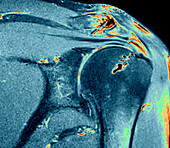
13600386 - Calcified tendinitis, MRI scan

12947677 - Dislocated shoulder, X-ray

14078634 - Subacromial bursitis of the shoulder, illustration
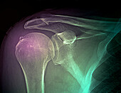
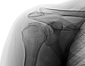
13600399 - Calcific tendinitis, X-ray
13443480 - Total hip joint prosthesis, illustration

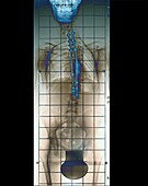
13426355 - Spinal decompression surgery
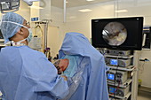
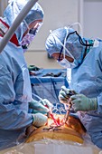
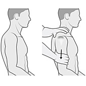
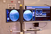
13272924 - Shoulder arthroscopy

12960957 - Spinal surgeons performing an operation
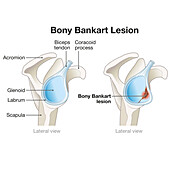
14078558 - Bony bankart lesion in the shoulder, illustration

13732382 - Human cartilage, light micrograph
13600838 - Osteoarthritis of the hip, X-ray
13474193 - Cross-section of chest bones, illustration
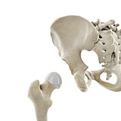
13444328 - Dislocated hip joint, illustration

13426354 - Spinal decompression surgery
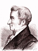
13258076 - Jacques Mathieu Delpech, French surgeon
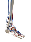
13224218 - Blood vessels of the foot, illustration
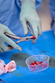
12960981 - Bone graft preparation during spinal surgery
13600709 - Foot bones, illustration
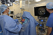
13272920 - Shoulder arthroscopy
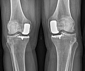
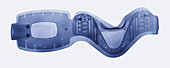
13218422 - Partial knee replacement, X-ray

14078604 - Shoulder examination, illustration
14078603 - Shoulder examination, illustration
14078601 - Shoulder examination, illustration
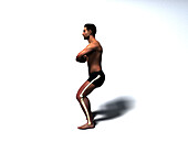
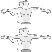
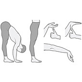
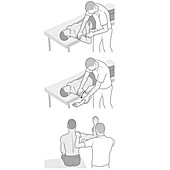
13619670 - Man squatting, illustration
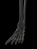
13600708 - Foot bones, illustration
13426362 - Patient in surgery
13224527 - Ligaments of the hip, illustration
12960959 - Spinal surgeons performing an operation

12960944 - Spinal surgeons during an operation
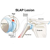
14078626 - SLAP lesion of the shoulder, illustration
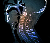
13600167 - Odontoid process fracture, CT scan
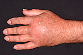
13295917 - Swelling due to fibrous dysplasia
13224514 - Ligaments of the foot, illustration
14078627 - SLAP lesion paralabral cyst in the shoulder, illustration
14078611 - Shoulder examination, illustration
13600755 - Foot bones, illustration
13600166 - Odontoid process fracture, CT scan
13600163 - Odontoid process fracture, CT scan
13218282 - Spondylolisthesis, X-ray

12991819 - Limb splints, 19th and 20th century
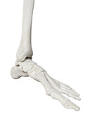
13600801 - Foot bones, illustration
13585361 - Gout
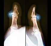
13453244 - Pseudogout of a thumb, X-ray scans
13426359 - Imaging during spinal decompression surgery

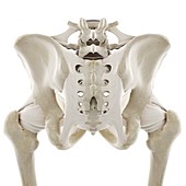
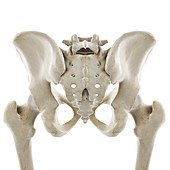
13600703 - Hip bones, illustration
13600397 - Calcific tendinitis, X-ray
13452945 - Harrington rod spinal implants in scoliosis, X-ray

13426361 - Patient in surgery
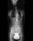
13355808 - Neck brace, X-ray

13224249 - Bones of the foot, illustration
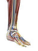
13224192 - Anatomy of the foot, illustration

13218421 - Partial knee replacement, X-ray

12960967 - Spinal surgeons performing an operation

12960966 - Spinal surgeons performing an operation
13732375 - Human cartilage, light micrograph

13471957 - Human sphenoid bone, illustration
13452943 - Harrington rod spinal implants in scoliosis, X-ray

12960946 - Spinal surgeons conferring during an operation

13600396 - Calcified tendinitis, MRI scan
13224511 - Ligaments of the foot, illustration
13224405 - Human hip bone, illustration
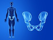
13224353 - Hip, illustration
13224348 - Hip joint, illustration
nächste Seite
Orthopädie Bilder ❘ Science Photo Library