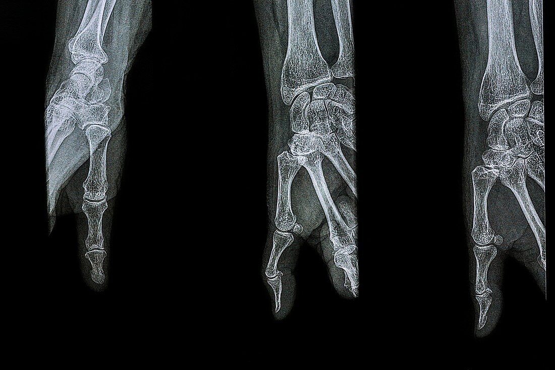
Diagnostic thumb X-rays
Bildnummer 11703480

| Diagnostic thumb X-rays. Three X-rays of a human thumb from different perspectives to enable diagnosis of medical conditions. At left is the thumb from above,with the wrist rotated so that the palm is facing downwards. At centre is the thumb sideways on,with the hand facing upwards and the fingers clenched into the palm. At right is the thumb with the hand in the same position,but with the fingers extended. The thumb consists of a metacarpal bone and two phalanx bones. The two images at centre and right show sesamoid bones (small,cubical) embedded within tendons in the joint between the metacarpal and first phalanx (the metacarpophalangeal joint) | |
| Lizenzart: | Lizenzpflichtig |
| Credit: | Science Photo Library / Gadsby, Brian |
| Bildgröße: | 5134 px × 3423 px |
| Modell-Rechte: | nicht erforderlich |
| Eigentums-Rechte: | nicht erforderlich |
| Restrictions: | - |
Preise für dieses Bild ab 15 €
Universitäten & Organisationen
(Informationsmaterial Digital, Informationsmaterial Print, Lehrmaterial Digital etc.)
ab 15 €
Redaktionell
(Bücher, Bücher: Sach- und Fachliteratur, Digitale Medien (redaktionell) etc.)
ab 30 €
Werbung
(Anzeigen, Aussenwerbung, Digitale Medien, Fernsehwerbung, Karten, Werbemittel, Zeitschriften etc.)
ab 55 €
Handelsprodukte
(bedruckte Textilie, Kalender, Postkarte, Grußkarte, Verpackung etc.)
ab 75 €
Pauschalpreise
Rechtepakete für die unbeschränkte Bildnutzung in Print oder Online
ab 495 €
Keywords
- 3,
- Anatomie,
- anatomisch,
- Artikulation,
- Close-up,
- Daumen,
- Detail,
- Diagnose,
- dorsal,
- Drei,
- Einfarbig,
- Gelenk,
- Gelenke,
- gesund,
- Hand,
- Handgelenk,
- Joint,
- Knochen,
- medizinisch,
- menschlicher Körper,
- Mittelhandknochen,
- Niemand,
- normal,
- Orthopädie,
- Phalangen,
- Phalanx,
- Radiographie,
- Röntgen,
- Röntgengerät,
- Schwarz und weiß,
- schwarzer Hintergrund,
- Seite,
- seitlich,
- Skelett,
- Skelett-,
- Trio
