Platelet, SEM
Bildnummer 13733305
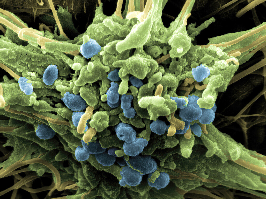
| Coloured scanning electron micrograph (SEM) showing an activated platelet (thrombocyte) trapped in a mesh of fibrin strands (yellow) during the hemostatic process of wound healing. The shell (green) of the platelet has collapsed and numerous vesicles (blue) have been released into the wound area. The vesicles contain a large number of enzymes and growth factors, thereby attracting cells needed during the following stages of wound healing. Vessel wall injury triggers the attachment and activation of platelets and the generation of fibrin polymers from fibrinogen. Platelets and fibrin combine to form a blood clot containing erythrocytes (red blood cells). Magnification: ×17000 when printed at 10 centimetres wide. | |
| Lizenzart: | Lizenzpflichtig |
| Credit: | Science Photo Library / PETER SCHUPBACH |
| Bildgröße: | 4096 px × 3072 px |
| Modell-Rechte: | nicht erforderlich |
| Eigentums-Rechte: | nicht erforderlich |
| Restrictions: | - |
Preise für dieses Bild ab 15 €
Universitäten & Organisationen
(Informationsmaterial Digital, Informationsmaterial Print, Lehrmaterial Digital etc.)
ab 15 €
Redaktionell
(Bücher, Bücher: Sach- und Fachliteratur, Digitale Medien (redaktionell) etc.)
ab 30 €
Werbung
(Anzeigen, Aussenwerbung, Digitale Medien, Fernsehwerbung, Karten, Werbemittel, Zeitschriften etc.)
ab 55 €
Handelsprodukte
(bedruckte Textilie, Kalender, Postkarte, Grußkarte, Verpackung etc.)
ab 75 €
Pauschalpreise
Rechtepakete für die unbeschränkte Bildnutzung in Print oder Online
ab 495 €
Keywords
- Anatomie,
- anatomisch,
- Biologie,
- biologisch,
- Blut,
- Blutplättchen,
- Blutzellen,
- farbig,
- gefärbt,
- Gerinnsel,
- Gerinnung,
- Gesundheit,
- Gesundheitswesen,
- Granulat,
- Hämatologie,
- hämatologisch,
- Medizin,
- Mensch,
- Niemand,
- Physiologie,
- physiologisch,
- Rasterelektronenmikroskop,
- rasterelektronenmikroskopische Aufnahme,
- REM,
- thrombozyten,
- Thrombus,
- Verletzung,
- Wunde,
- Wundheilung