Liver ischemia, light micrograph
Bildnummer 13732411
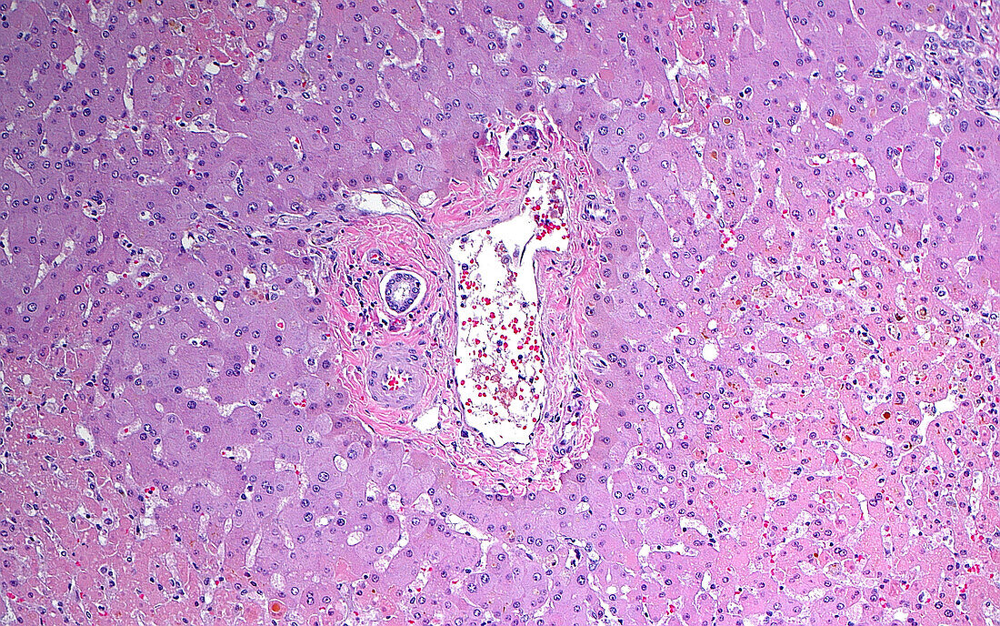
| Light micrograph of a centrilobular pattern of necrotic liver cells. The necrotic liver cells are the lighter pink cells mostly in the bottom left and bottom right corners of the picture. In the centre of the image is a portal tract consisting of an artery, vein, and bile duct. The liver cells around the portal tract are not necrotic because they receive an adequate supply of blood from the nearby vessels. The liver cells further out are necrotic, indicating ischemia or a lack of enough oxygen and nutrients further away from the blood source. Such a pattern of necrosis can occur when there are other factors in the body that decrease the availability of oxygen or nutrients in the blood, such as infection or shock. Haematoxylin and eosin stained tissue section. Magnification: 100x when printed at 10 cm. | |
| Lizenzart: | Lizenzpflichtig |
| Credit: | Science Photo Library / ZIAD M. EL-ZAATARI |
| Bildgröße: | 5000 px × 3125 px |
| Modell-Rechte: | nicht erforderlich |
| Eigentums-Rechte: | nicht erforderlich |
| Restrictions: | - |
Preise für dieses Bild ab 15 €
Universitäten & Organisationen
(Informationsmaterial Digital, Informationsmaterial Print, Lehrmaterial Digital etc.)
ab 15 €
Redaktionell
(Bücher, Bücher: Sach- und Fachliteratur, Digitale Medien (redaktionell) etc.)
ab 30 €
Werbung
(Anzeigen, Aussenwerbung, Digitale Medien, Fernsehwerbung, Karten, Werbemittel, Zeitschriften etc.)
ab 55 €
Handelsprodukte
(bedruckte Textilie, Kalender, Postkarte, Grußkarte, Verpackung etc.)
ab 75 €
Pauschalpreise
Rechtepakete für die unbeschränkte Bildnutzung in Print oder Online
ab 495 €