Development of retinal cancer, illustration
Bildnummer 12971400
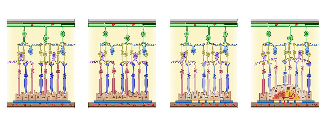
| Development of retinal cancer, illustration. Sequence of four images of the cellular structure and layers of the retina, the light-sensitive layer lining the inside of the back of the eye. Running from left to right, the images show the development of a tumour (growth) on the retina, originating from the choroid (the layer across bottom). Above this layer is Bruch's membrane, and the retinal pigment epithelium (RPE). Above these are the photoreceptor cells, the nuclear layers, and the ganglion layer (green) and nerve fibre layer (top). Cancers affecting the retina usually occur in the choroid, a dense layer of blood vessels that supplies the retina. | |
| Lizenzart: | Lizenzpflichtig |
| Credit: | Science Photo Library / De Angelis, Maurizio |
| Bildgröße: | 5240 px × 2001 px |
| Modell-Rechte: | nicht erforderlich |
| Eigentums-Rechte: | nicht erforderlich |
| Restrictions: | - |
Preise für dieses Bild ab 15 €
Universitäten & Organisationen
(Informationsmaterial Digital, Informationsmaterial Print, Lehrmaterial Digital etc.)
ab 15 €
Redaktionell
(Bücher, Bücher: Sach- und Fachliteratur, Digitale Medien (redaktionell) etc.)
ab 30 €
Werbung
(Anzeigen, Aussenwerbung, Digitale Medien, Fernsehwerbung, Karten, Werbemittel, Zeitschriften etc.)
ab 55 €
Handelsprodukte
(bedruckte Textilie, Kalender, Postkarte, Grußkarte, Verpackung etc.)
ab 75 €
Pauschalpreise
Rechtepakete für die unbeschränkte Bildnutzung in Print oder Online
ab 495 €
Keywords
- abnormal,
- Aderhaut,
- Auge,
- Augen-,
- Augenheilkunde,
- ausgeschnitten,
- Ausschnitte,
- Entwicklung,
- Ganglion,
- Illustration,
- Kondition,
- Krankheit,
- Krebs,
- krebsartig,
- Kunstwerk,
- maligne,
- Malignom,
- Medizin,
- medizinisch,
- menschlicher Körper,
- Netzhaut-,
- Niemand,
- okular,
- Reihenfolge,
- Retina,
- retinales Pigmentepithel,
- rpe,
- Schichten,
- Serie,
- Störung,
- Tumor,
- ungesund,
- wachsend,
- Wachstum,
- weißer Hintergrund