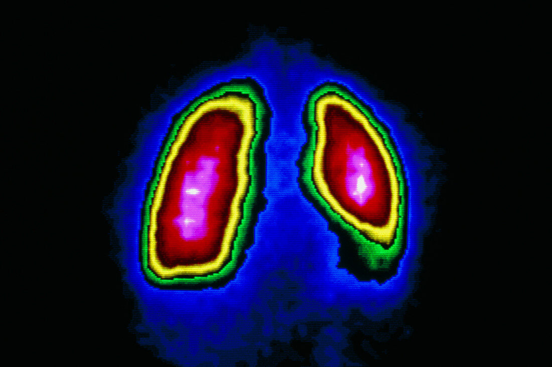
F/colour gamma scan of healthy human lungs
Bildnummer 12968910

| Human lungs. False-colour computer enhanced gamma scan (scintigram) of healthy human lungs. The lungs appear inflated. The central region of the lungs (red) has absorbed more radioactive tracer than the outer regions (yellow,green). Gamma scanning involves introducing a radioactive iso- tope into the body,in this case Technetium detected by a gamma camera. The isotope emits gamma rays which are detected as flashes of light. Isotopes are chosen for their ability to gather in cancerous or inflamed tissue,producing hot spots"" that would be highlighted on the scan. Gamma scan sections of lung tissue can be examined in slices." | |
| Lizenzart: | Lizenzpflichtig |
| Credit: | Science Photo Library / CENTRE JEAN PERRIN |
| Bildgröße: | 4594 px × 3052 px |
| Modell-Rechte: | nicht erforderlich |
| Eigentums-Rechte: | nicht erforderlich |
| Restrictions: | - |
Preise für dieses Bild ab 15 €
Universitäten & Organisationen
(Informationsmaterial Digital, Informationsmaterial Print, Lehrmaterial Digital etc.)
ab 15 €
Redaktionell
(Bücher, Bücher: Sach- und Fachliteratur, Digitale Medien (redaktionell) etc.)
ab 30 €
Werbung
(Anzeigen, Aussenwerbung, Digitale Medien, Fernsehwerbung, Karten, Werbemittel, Zeitschriften etc.)
ab 55 €
Handelsprodukte
(bedruckte Textilie, Kalender, Postkarte, Grußkarte, Verpackung etc.)
ab 75 €
Pauschalpreise
Rechtepakete für die unbeschränkte Bildnutzung in Print oder Online
ab 495 €
