Multimodal cancer imaging with nanoparticles
Bildnummer 12638798
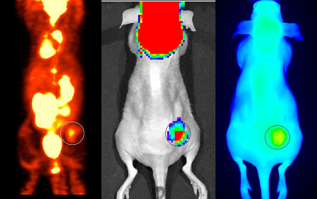
| Multimodal cancer imaging with nanoparticles. Three medical imaging methods being used to study nanoparticles developed to treat cancers in mouse models. Here, therapeutic nanoparticles tagged with a fluorescent molecule have accumulated in a mouse's tumour (yellow, circled at right). Simultaneously, luminescent 18F-fluorodeoxyglucose (FDG) Cerenkov imaging was used to confirm the tumour location (circled, centre), with multicolours indicating different FDG absorption rates. FDG also accumulated in the mouse's brain. The tumour location was also confirmed by a positron emission tomography (PET) scan (circled, left). This montage was created in 2016, as part of research carried out at Washington University in St Louis, USA. | |
| Lizenzart: | Lizenzpflichtig |
| Credit: | Science Photo Library / Washington University in St Louis / NATIONAL CANCER INSTITUTE |
| Bildgröße: | 5253 px × 3300 px |
| Modell-Rechte: | nicht erforderlich |
| Eigentums-Rechte: | nicht erforderlich |
| Restrictions: | - |
Preise für dieses Bild ab 15 €
Universitäten & Organisationen
(Informationsmaterial Digital, Informationsmaterial Print, Lehrmaterial Digital etc.)
ab 15 €
Redaktionell
(Bücher, Bücher: Sach- und Fachliteratur, Digitale Medien (redaktionell) etc.)
ab 30 €
Werbung
(Anzeigen, Aussenwerbung, Digitale Medien, Fernsehwerbung, Karten, Werbemittel, Zeitschriften etc.)
ab 55 €
Handelsprodukte
(bedruckte Textilie, Kalender, Postkarte, Grußkarte, Verpackung etc.)
ab 75 €
Pauschalpreise
Rechtepakete für die unbeschränkte Bildnutzung in Print oder Online
ab 495 €
Keywords
- 2016,
- 21. Jahrhundert,
- 3,
- Amerikanisch,
- Arzneimittel,
- Behandlung,
- biologisch,
- Drei,
- farbig,
- Fluoreszenz,
- fluoreszierend,
- gefärbt,
- Haustier,
- Labor,
- Maus,
- Medizin,
- medizinisch,
- Molekül,
- Molekularbiologie,
- Nano,
- Nanopartikel,
- Nanotechnologie,
- Niemand,
- Onkologie,
- Scan,
- Scanner,
- therapeutisch,
- Tier,
- Trio,
- uns,
- Zelle