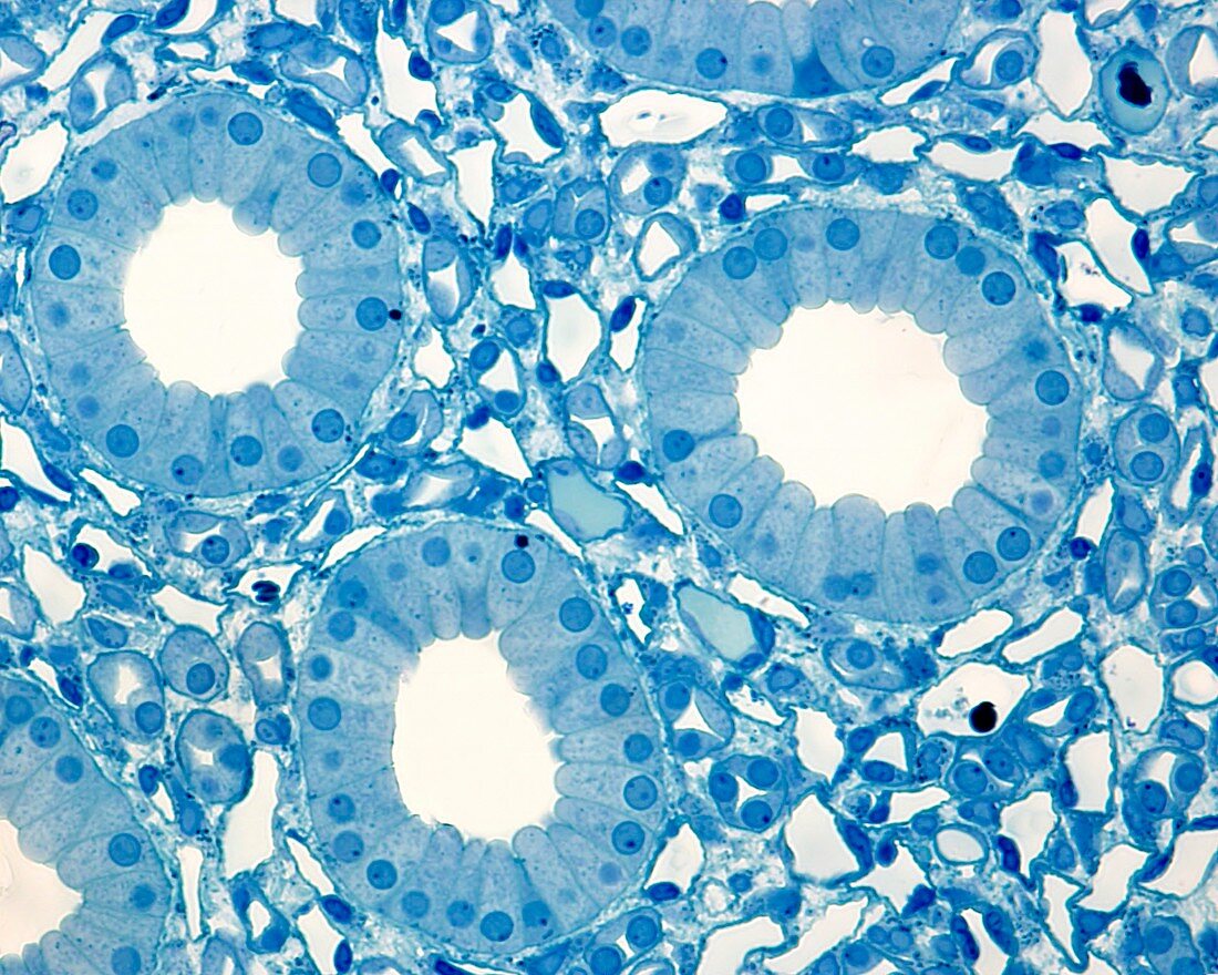
Renal medulla, light micrograph
Bildnummer 12360964

| Light micrograph of the inner region of the renal medulla. 0.5 micrometre thick section stained with toluidine blue and embedded in a synthetic resin. The micrograph shows three cross-sectioned papillary or Bellini ducts. These ducts, resulting from the fusion of collecting tubes (an average of 7 fusions) reach a diameter of 100-200 micrometres, and are covered by a simple columnar epithelium, formed by clear cells. The space between the ducts is occupied by very thin tubes of two types. One, the most abundant, is lined by a very thin simple squamous epithelium (endothelium). They are blood vessels, both capillaries and venules. The second type has a smaller diameter and has a higher epithelium: they are Henle's loops. Magnification: x360 when printed at 10 centimetres across. | |
| Lizenzart: | Lizenzpflichtig |
| Credit: | Science Photo Library / JOSE CALVO |
| Bildgröße: | 4674 px × 3739 px |
| Modell-Rechte: | nicht erforderlich |
| Eigentums-Rechte: | nicht erforderlich |
| Restrictions: | - |
Preise für dieses Bild ab 15 €
Universitäten & Organisationen
(Informationsmaterial Digital, Informationsmaterial Print, Lehrmaterial Digital etc.)
ab 15 €
Redaktionell
(Bücher, Bücher: Sach- und Fachliteratur, Digitale Medien (redaktionell) etc.)
ab 30 €
Werbung
(Anzeigen, Aussenwerbung, Digitale Medien, Fernsehwerbung, Karten, Werbemittel, Zeitschriften etc.)
ab 55 €
Handelsprodukte
(bedruckte Textilie, Kalender, Postkarte, Grußkarte, Verpackung etc.)
ab 75 €
Pauschalpreise
Rechtepakete für die unbeschränkte Bildnutzung in Print oder Online
ab 495 €
Keywords
- Adventitia,
- Anatomie,
- anatomisch,
- Ausscheidung,
- Bindegewebe,
- Biologie,
- biologisch,
- Blase,
- Blutgefäß,
- Blutgefäße,
- Bürstensaum,
- Epithel,
- epithelial,
- Epithelien,
- Erythrozyt,
- Erythrozyten,
- Gefäß,
- Gefäße,
- gesund,
- Gewebe,
- glomerulär,
- Glomerulus,
- Harnsystem,
- Henle-Schleife,
- Histologie,
- histologisch,
- kapillar,
- Lichtmikroskop,
- lichtmikroskopische Aufnahme,
- Lumen,
- Mark,
- Menschliche Biologie,
- menschlicher Körper,
- Mikrofotografie,
- Mikroskop,
- Mikroskopie,
- mikroskopisch,
- Mikrovilli,
- Mikrovillus,
- nephron,
- Niere,
- Nieren,
- Nieren-,
- Plattenepithel,
- Querschnitt,
- rot,
- rote Blutkörperchen,
- Schleimhaut,
- Submukosa,
- Übergangsepithel,
- Vergrößerung,
- Zelle,
- Zellen
