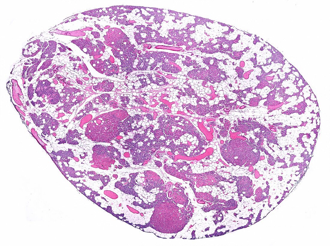
Parathyroid gland, light micrograph
Bildnummer 12360949

| Low magnification light micrograph showing a parathyroid gland of an aged human adult. Most of the volume of the gland corresponds to adipose tissue that has proliferated in the connective tissue septa. The parenchyma has been reduced, since the total volume of the gland hardly changes. In spite of the very low magnification, the large nodules formed by oxyphyl cells without endocrine function stand out in the parathyroid parenchyma. Magnification: x18 when printed at 10 centimetres across. | |
| Lizenzart: | Lizenzpflichtig |
| Credit: | Science Photo Library / JOSE CALVO |
| Bildgröße: | 6165 px × 4608 px |
| Modell-Rechte: | nicht erforderlich |
| Eigentums-Rechte: | nicht erforderlich |
| Restrictions: | - |
Preise für dieses Bild ab 15 €
Universitäten & Organisationen
(Informationsmaterial Digital, Informationsmaterial Print, Lehrmaterial Digital etc.)
ab 15 €
Redaktionell
(Bücher, Bücher: Sach- und Fachliteratur, Digitale Medien (redaktionell) etc.)
ab 30 €
Werbung
(Anzeigen, Aussenwerbung, Digitale Medien, Fernsehwerbung, Karten, Werbemittel, Zeitschriften etc.)
ab 55 €
Handelsprodukte
(bedruckte Textilie, Kalender, Postkarte, Grußkarte, Verpackung etc.)
ab 75 €
Pauschalpreise
Rechtepakete für die unbeschränkte Bildnutzung in Print oder Online
ab 495 €
Keywords
- Biologie,
- biologisch,
- Blutgefäße,
- Drüse,
- endokrin,
- Endokrinologie,
- Endokrinsystem,
- gesund,
- Hämatoxylin-Eosin-Färbung,
- Histologie,
- histologisch,
- hormonell,
- kapillar,
- Kerne,
- Lichtmikroskop,
- lichtmikroskopische Aufnahme,
- Menschliche Biologie,
- menschlicher Körper,
- Mikrofotografie,
- Mikroskopie,
- mikroskopisch,
- Zelle,
- Zellen,
- zellular
