Coloured SEM of foreparts of dust mite.
Bildnummer 12261728
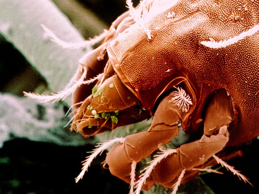
| Dust mite. Coloured scanning electron micrograph (SEM) of a dust mite, Glycyphagus domestica. The mite's body is in three parts: the gnathosoma (head region) and propodosma (carrying the first and second pairs of walking legs), both shown here, and the hysterosoma where the third and fourth pairs of walking legs are located. The dust mite thrives in damp houses, feeding on groceries, furniture stuffing, wallpaper paste and other such delicacies. Magnification: x280 at 5x7cm size. x740 at 8x6, x395 at 10x7cm master size | |
| Lizenzart: | Lizenzpflichtig |
| Credit: | Science Photo Library / Power And Syred |
| Bildgröße: | 4897 px × 3661 px |
| Modell-Rechte: | nicht erforderlich |
| Eigentums-Rechte: | nicht erforderlich |
| Restrictions: | - |
Preise für dieses Bild ab 15 €
Universitäten & Organisationen
(Informationsmaterial Digital, Informationsmaterial Print, Lehrmaterial Digital etc.)
ab 15 €
Redaktionell
(Bücher, Bücher: Sach- und Fachliteratur, Digitale Medien (redaktionell) etc.)
ab 30 €
Werbung
(Anzeigen, Aussenwerbung, Digitale Medien, Fernsehwerbung, Karten, Werbemittel, Zeitschriften etc.)
ab 55 €
Handelsprodukte
(bedruckte Textilie, Kalender, Postkarte, Grußkarte, Verpackung etc.)
ab 75 €
Pauschalpreise
Rechtepakete für die unbeschränkte Bildnutzung in Print oder Online
ab 495 €