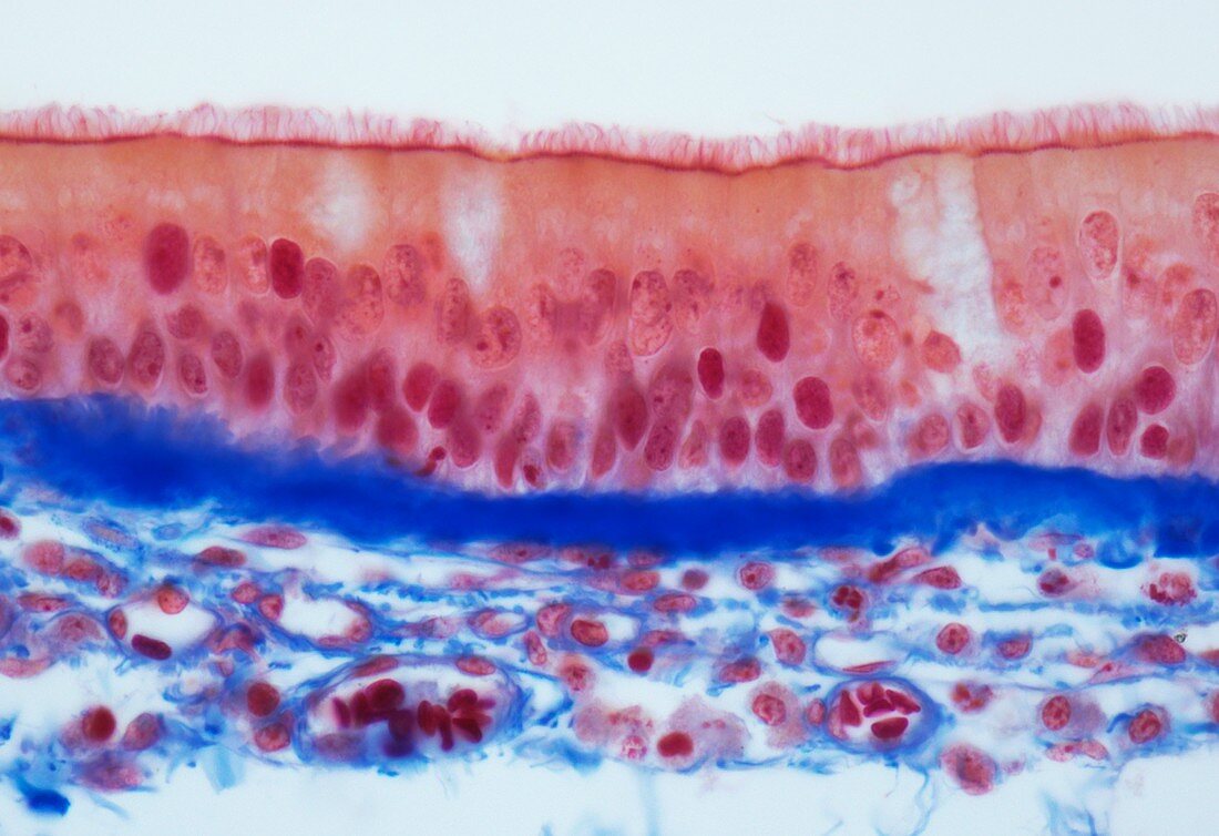
Tracheal epithelium,LM
Bildnummer 12098243

| Tracheal epithelium. Light micrograph (LM) of a vertical section through the pseudostratified columnar epithelium from the trachea,the tube in the throat leading from the mouth to the bronchus and the lungs. Cilia are seen across the inner surface (top). These help to move mucus along the trachea from the lungs to the mouth,removing dust,cellular debris and pathogens. Below the cilia are the columnar cells of the tracheal epithelium (pink),with their nuclei at their bases. Below those two layers is a layer of connective tissue (blue) and blood vessels (red). Magnification: x3300 when printed 10 centimetres wide | |
| Lizenzart: | Lizenzfrei |
| Credit: | Science Photo Library / Gschmeissner, Steve |
| Modell-Rechte: | nicht erforderlich |
| Eigentums-Rechte: | nicht erforderlich |
| Restrictions: | - |
Preise für dieses Bild ab 29 €
Für digitale Nutzung (72 dpi)
ab 29 €
Für Druckauflösung (300 dpi)
ab 300 €
Keywords
- Anatomie,
- anatomisch,
- Atmungssystem,
- Bindegewebe,
- Biologie,
- biologisch,
- Blutgefäß,
- Blutgefäße,
- Epithel,
- epithelial,
- Gefäße,
- gesund,
- Gewebe,
- Histologie,
- histologisch,
- Kehle,
- Kerne,
- Lichtmikroskop,
- lichtmikroskopische Aufnahme,
- Luftröhre,
- Medizin,
- menschlicher Körper,
- Niemand,
- normal,
- pulmonal,
- Schleimtransport,
- Sektion,
- sektioniert,
- Trachealepithel,
- Wimpern,
- Zelle
