Pericytes,SEM
Bildnummer 12047431

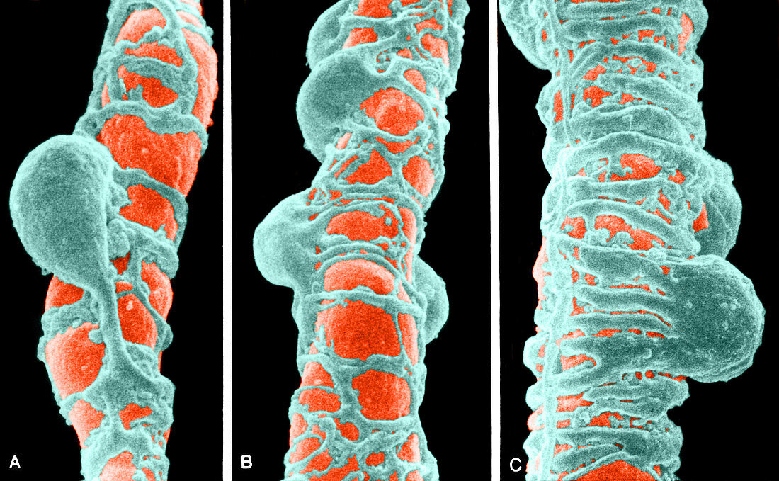
| Colour enhanced scanning electron micrograph of small blood vessels showing pericytes with processes encircling the vessel wall. Image A shows a pericyte of a capillary with primary processes directed longitudinally and secondary processes running circumferentially. Image B shows an arterial capillary with numerous associated pericytes. Image C shows a terminal arteriole with pericyte and circular smooth muscle cells. Magnification unknown | |
| Lizenzart: | Lizenzpflichtig |
| Credit: | Science Photo Library / Fawcett, Don W. |
| Bildgröße: | 4789 px × 2954 px |
| Modell-Rechte: | nicht erforderlich |
| Eigentums-Rechte: | nicht erforderlich |
| Restrictions: |
|
Preise für dieses Bild ab 15 €
Universitäten & Organisationen
(Informationsmaterial Digital, Informationsmaterial Print, Lehrmaterial Digital etc.)
ab 15 €
Redaktionell
(Bücher, Bücher: Sach- und Fachliteratur, Digitale Medien (redaktionell) etc.)
ab 30 €
Werbung
(Anzeigen, Aussenwerbung, Digitale Medien, Fernsehwerbung, Karten, Werbemittel, Zeitschriften etc.)
ab 55 €
Handelsprodukte
(bedruckte Textilie, Kalender, Postkarte, Grußkarte, Verpackung etc.)
ab 75 €
Pauschalpreise
Rechtepakete für die unbeschränkte Bildnutzung in Print oder Online
ab 495 €
Keywords
- arteriell,
- arterielle Kapillare,
- Bindegewebe,
- Blut,
- Blutgefäß,
- eingefärbt,
- Elektronenmikroskopie,
- elektronenmikroskopische Aufnahme,
- Gefäß,
- Gewebe,
- Histologie,
- kapillar,
- Kreislauf,
- Mikrofotografie,
- Mikrographie,
- Niemand,
- Rasterelektronenmikroskopie,
- rasterelektronenmikroskopische Aufnahme,
- REM,
- verbessert,
- Zelle,
- Zytologie
