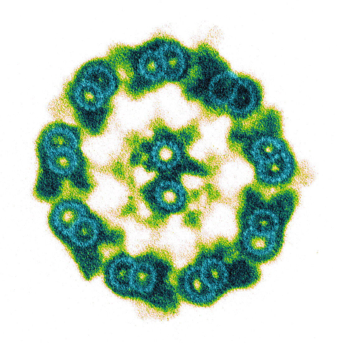
Axoneme of Tetrahymena thermophila
Bildnummer 12046594

| Colour enhanced transmission electron micrograph of the cilia of Tetrahymena thermophila are used not only for feeding and propelling the cell through the medium but also for sensing the environment around them. This image is of an isolated axoneme with the membrane removed and stained with tannic acid. The tannic acid allows the visualization of the individual tubulin heterodimers that make up the walls of the microtubule doublets. The central pair and A tubules of the microtubule doublets are made up of 13 tubulin heterodimers whereas the B tubules of the microtubule doublets are made up of 11 heterodimers. Magnification 125,000X | |
| Lizenzart: | Lizenzpflichtig |
| Credit: | Science Photo Library / Bell, Aaron J. |
| Bildgröße: | 3000 px × 3047 px |
| Modell-Rechte: | nicht erforderlich |
| Eigentums-Rechte: | nicht erforderlich |
| Restrictions: |
|
Preise für dieses Bild ab 15 €
Universitäten & Organisationen
(Informationsmaterial Digital, Informationsmaterial Print, Lehrmaterial Digital etc.)
ab 15 €
Redaktionell
(Bücher, Bücher: Sach- und Fachliteratur, Digitale Medien (redaktionell) etc.)
ab 30 €
Werbung
(Anzeigen, Aussenwerbung, Digitale Medien, Fernsehwerbung, Karten, Werbemittel, Zeitschriften etc.)
ab 55 €
Handelsprodukte
(bedruckte Textilie, Kalender, Postkarte, Grußkarte, Verpackung etc.)
ab 75 €
Pauschalpreise
Rechtepakete für die unbeschränkte Bildnutzung in Print oder Online
ab 495 €
