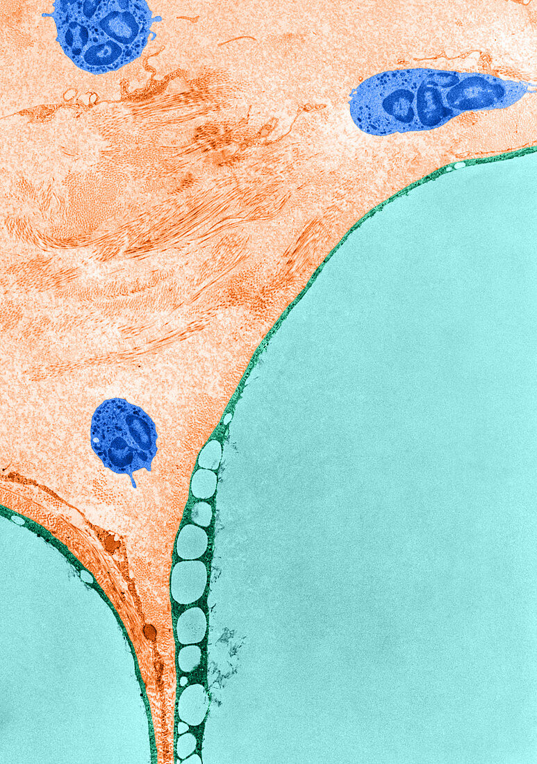
TEM of Adipose Cells
Bildnummer 12046441

| Colour enhanced transmission electron micrograph of portions of two adipose cells and associated connective tissue from the epididymal fat pad of a rat. Adipose cells are among the largest cells in the body,but less than one fortieth of their total volume is metabolically active. The nucleus is displaced to the periphery and the cytoplasm is reduced to a thin layer surrounding a large globule of accumulated lipid. The very large relative size of adipose cells can be appreciated in the accompanying micrograph by comparison of three leucocytes in the field with portions of two fat cells included in the lower half of the figure. Newly synthesized lipid first forms small droplets in the peripheral layer of cytoplasm (at arrows) and these subsequently coalesce with the large central lipid drop | |
| Lizenzart: | Lizenzpflichtig |
| Credit: | Science Photo Library / Fawcett, Don W. |
| Bildgröße: | 3347 px × 4762 px |
| Modell-Rechte: | nicht erforderlich |
| Eigentums-Rechte: | nicht erforderlich |
| Restrictions: |
|
Preise für dieses Bild ab 15 €
Universitäten & Organisationen
(Informationsmaterial Digital, Informationsmaterial Print, Lehrmaterial Digital etc.)
ab 15 €
Redaktionell
(Bücher, Bücher: Sach- und Fachliteratur, Digitale Medien (redaktionell) etc.)
ab 30 €
Werbung
(Anzeigen, Aussenwerbung, Digitale Medien, Fernsehwerbung, Karten, Werbemittel, Zeitschriften etc.)
ab 55 €
Handelsprodukte
(bedruckte Textilie, Kalender, Postkarte, Grußkarte, Verpackung etc.)
ab 75 €
Pauschalpreise
Rechtepakete für die unbeschränkte Bildnutzung in Print oder Online
ab 495 €
