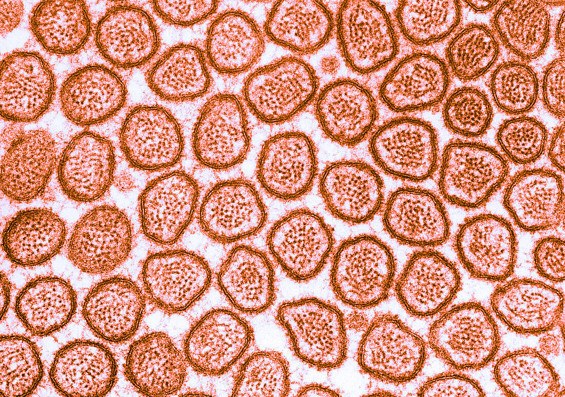
Microvilli in Intestinal Epithelium,TEM
Bildnummer 12046322

| Colour enhanced transmission electron micrograph of microvilli of the brush border of cat intestinal epithelium. This transverse section of intestinal brush border shows the actin filaments that form the core of each microvillus. At the villus tip,the relation of the filaments to the membrane is obscured by a layer of dense amorphous material associated with the inner aspect of the membrane | |
| Lizenzart: | Lizenzpflichtig |
| Credit: | Science Photo Library / Fawcett, Don W. |
| Bildgröße: | 4437 px × 3119 px |
| Modell-Rechte: | nicht erforderlich |
| Eigentums-Rechte: | nicht erforderlich |
| Restrictions: |
|
Preise für dieses Bild ab 15 €
Universitäten & Organisationen
(Informationsmaterial Digital, Informationsmaterial Print, Lehrmaterial Digital etc.)
ab 15 €
Redaktionell
(Bücher, Bücher: Sach- und Fachliteratur, Digitale Medien (redaktionell) etc.)
ab 30 €
Werbung
(Anzeigen, Aussenwerbung, Digitale Medien, Fernsehwerbung, Karten, Werbemittel, Zeitschriften etc.)
ab 55 €
Handelsprodukte
(bedruckte Textilie, Kalender, Postkarte, Grußkarte, Verpackung etc.)
ab 75 €
Pauschalpreise
Rechtepakete für die unbeschränkte Bildnutzung in Print oder Online
ab 495 €
