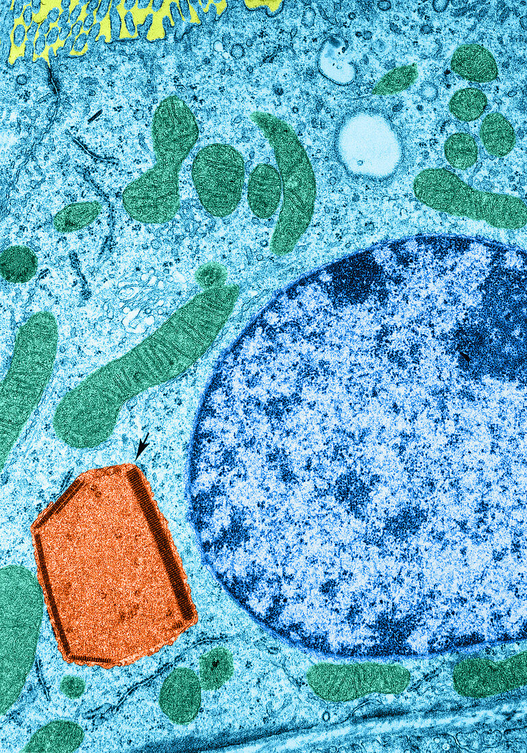
Lysosomes,TEM
Bildnummer 12046189

| Colour enhanced transmission electron micrograph of a proximal convoluted tubule cell from a rat. A peroxisome (orange) is visible near the cell base containing both crystals and a few circular profiles of cylinders in cross section. Despite their unusual appearance,these organelles can be identified as peroxisomes by positive staining for catalase and a negative reaction for acid phosphatase. Micrograph courtesy of Michael Barrett and Paul Heidger. THE CELL 2nd edition | |
| Lizenzart: | Lizenzpflichtig |
| Credit: | Science Photo Library / Fawcett / Barett / Heidger |
| Bildgröße: | 3515 px × 5027 px |
| Modell-Rechte: | nicht erforderlich |
| Eigentums-Rechte: | nicht erforderlich |
| Restrictions: |
|
Preise für dieses Bild ab 15 €
Universitäten & Organisationen
(Informationsmaterial Digital, Informationsmaterial Print, Lehrmaterial Digital etc.)
ab 15 €
Redaktionell
(Bücher, Bücher: Sach- und Fachliteratur, Digitale Medien (redaktionell) etc.)
ab 30 €
Werbung
(Anzeigen, Aussenwerbung, Digitale Medien, Fernsehwerbung, Karten, Werbemittel, Zeitschriften etc.)
ab 55 €
Handelsprodukte
(bedruckte Textilie, Kalender, Postkarte, Grußkarte, Verpackung etc.)
ab 75 €
Pauschalpreise
Rechtepakete für die unbeschränkte Bildnutzung in Print oder Online
ab 495 €
