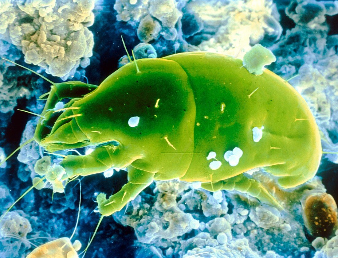
False-colour SEM of an unidentified dust mite
Bildnummer 11911008

| False-colour scanning electron micrograph of a an unidentified dust mite,in a sample of house-hold dust. Every house contains millions of such dust mites,living in the carpets and furniture and feeding on the flakes of skin shed by their human landlords. It is estimated that the average double bed contains about two million dust mites of various species. The body of the mite is divided into three distinct areas; the gnathosoma (head region,left),the propodosma (the second section which carries the first and second pairs of walking legs) and the hysterosoma (carrying third and fourth pairs of walking legs). Magnification: x240 at 6x7 cm size | |
| Lizenzart: | Lizenzpflichtig |
| Credit: | Science Photo Library / Photo Insolite Realite |
| Bildgröße: | 4838 px × 3697 px |
| Modell-Rechte: | nicht erforderlich |
| Eigentums-Rechte: | nicht erforderlich |
| Restrictions: | - |
Preise für dieses Bild ab 15 €
Universitäten & Organisationen
(Informationsmaterial Digital, Informationsmaterial Print, Lehrmaterial Digital etc.)
ab 15 €
Redaktionell
(Bücher, Bücher: Sach- und Fachliteratur, Digitale Medien (redaktionell) etc.)
ab 30 €
Werbung
(Anzeigen, Aussenwerbung, Digitale Medien, Fernsehwerbung, Karten, Werbemittel, Zeitschriften etc.)
ab 55 €
Handelsprodukte
(bedruckte Textilie, Kalender, Postkarte, Grußkarte, Verpackung etc.)
ab 75 €
Pauschalpreise
Rechtepakete für die unbeschränkte Bildnutzung in Print oder Online
ab 495 €
