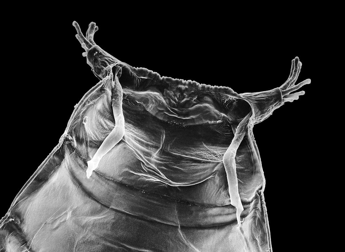
SEM of pupa case of fruit fly,Drosophila
Bildnummer 11908385

| Scanning electron micrograph of the inner surface of a pupal case,or puparium,of the fruit fly Drosophila melanogaster (wild type Oregon R). The view is of the anterior end showing the pair of anterior spiracles,or breathing tubes (horns),which are part of the respiratory apparatus for the pupa. The spiracles project out from the pupal wall,ending in tubelike extensions called spiracular papillae,which take in air through slits at their tips. Air is carried by tracheal tubes; the two main lateral branches are seen here where they pass into the spiracles. The apparatus is shed with the puparium. Magnification X200 (at 10x8 size) | |
| Lizenzart: | Lizenzpflichtig |
| Credit: | Science Photo Library / Burgess, Dr. Jeremy |
| Bildgröße: | 4961 px × 3620 px |
| Modell-Rechte: | nicht erforderlich |
| Eigentums-Rechte: | nicht erforderlich |
| Restrictions: | - |
Preise für dieses Bild ab 15 €
Universitäten & Organisationen
(Informationsmaterial Digital, Informationsmaterial Print, Lehrmaterial Digital etc.)
ab 15 €
Redaktionell
(Bücher, Bücher: Sach- und Fachliteratur, Digitale Medien (redaktionell) etc.)
ab 30 €
Werbung
(Anzeigen, Aussenwerbung, Digitale Medien, Fernsehwerbung, Karten, Werbemittel, Zeitschriften etc.)
ab 55 €
Handelsprodukte
(bedruckte Textilie, Kalender, Postkarte, Grußkarte, Verpackung etc.)
ab 75 €
Pauschalpreise
Rechtepakete für die unbeschränkte Bildnutzung in Print oder Online
ab 495 €
