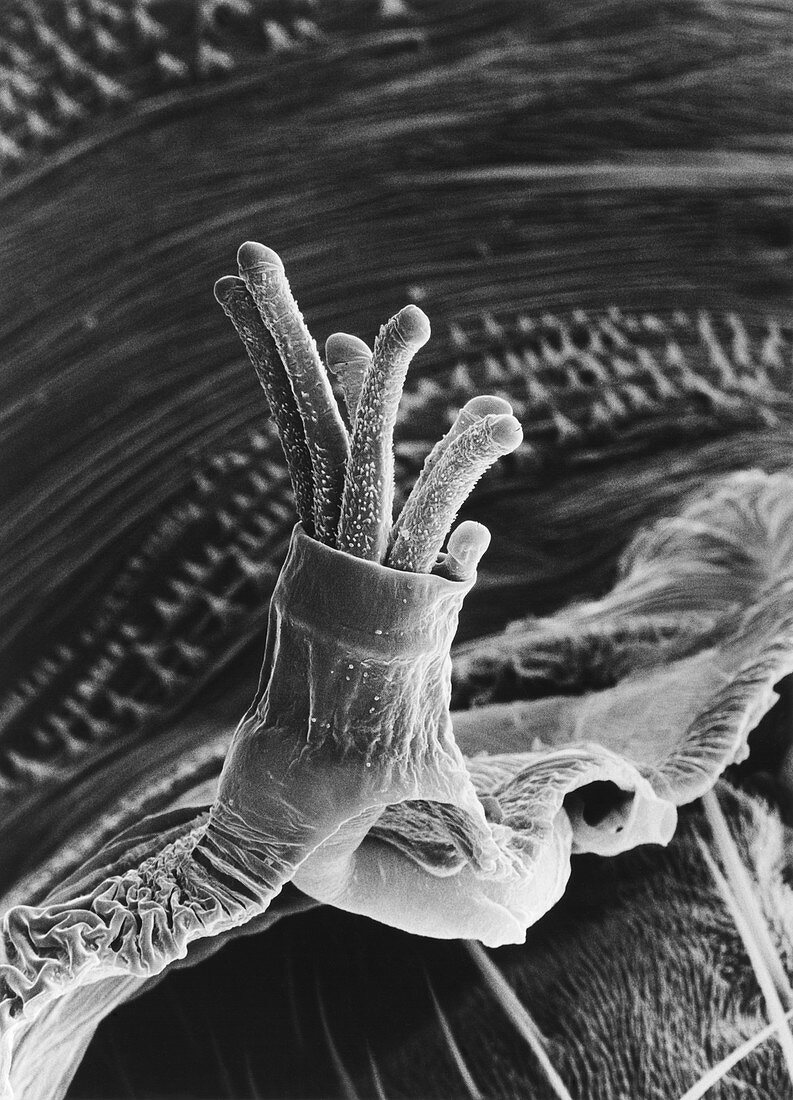
SEM of spiracle on pupal of fruit fly
Bildnummer 11908379

| Scanning electron micrograph of a spiracle,or breathing pore,on the pupal casing of the fruit fly Drosophila melanogaster. This anterior spiracle,one of two,draws air into the puparium. When the adult hatches the spiracles are shed with the pupal casing,as seen here. Air is drawn in through slits in the heads of the tubular extensions,called spiracular papillae. They feed into a pipe-like structure or tracheal tube,which conveys the air around the puparium. The trachea is spirally constructed & lined with chitin,a polysaccharide lending strength & resistance. Magnification: x520 at 8x10-inch size | |
| Lizenzart: | Lizenzpflichtig |
| Credit: | Science Photo Library / Burgess, Dr. Jeremy |
| Bildgröße: | 3576 px × 4961 px |
| Modell-Rechte: | nicht erforderlich |
| Eigentums-Rechte: | nicht erforderlich |
| Restrictions: | - |
Preise für dieses Bild ab 15 €
Universitäten & Organisationen
(Informationsmaterial Digital, Informationsmaterial Print, Lehrmaterial Digital etc.)
ab 15 €
Redaktionell
(Bücher, Bücher: Sach- und Fachliteratur, Digitale Medien (redaktionell) etc.)
ab 30 €
Werbung
(Anzeigen, Aussenwerbung, Digitale Medien, Fernsehwerbung, Karten, Werbemittel, Zeitschriften etc.)
ab 55 €
Handelsprodukte
(bedruckte Textilie, Kalender, Postkarte, Grußkarte, Verpackung etc.)
ab 75 €
Pauschalpreise
Rechtepakete für die unbeschränkte Bildnutzung in Print oder Online
ab 495 €
