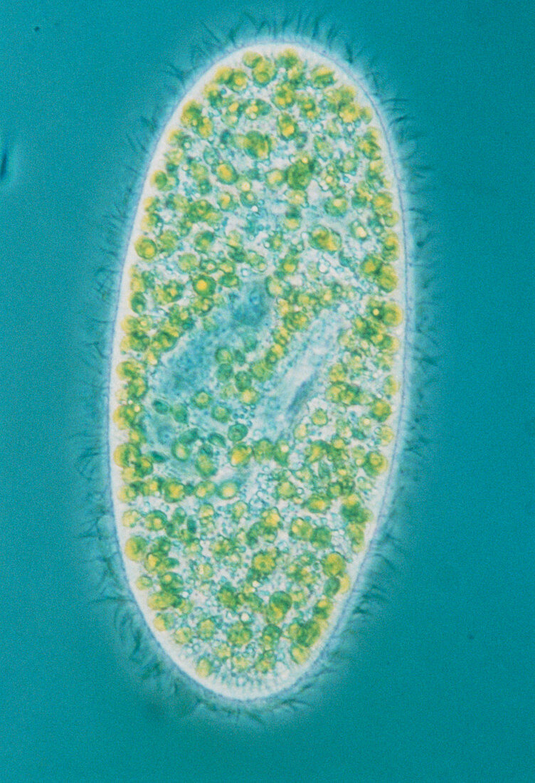
Light micrograph of Paramecium sp
Bildnummer 11905727

| Light micrograph of a ciliate protozoan Paramecium sp. The micrograph is one in a series of four images illustrating the different illumination systems of a light microscope. This is an example of phase contrast optics,which reduces colour saturation,but increases the amount of detail visible within the cytoplasm of the cell. The fringe of cilia,or fine hairs surrounding the organism,is visible using this technique. Magnification: x150 at 35mm size. Microcosmos P.192 Technical Appendix third of four (bottom left) Technical info: phase contrast optics | |
| Lizenzart: | Lizenzpflichtig |
| Credit: | Science Photo Library / Patterson, Dr. David J. |
| Bildgröße: | 3543 px × 5183 px |
| Modell-Rechte: | nicht erforderlich |
| Eigentums-Rechte: | nicht erforderlich |
| Restrictions: | - |
Preise für dieses Bild ab 15 €
Universitäten & Organisationen
(Informationsmaterial Digital, Informationsmaterial Print, Lehrmaterial Digital etc.)
ab 15 €
Redaktionell
(Bücher, Bücher: Sach- und Fachliteratur, Digitale Medien (redaktionell) etc.)
ab 30 €
Werbung
(Anzeigen, Aussenwerbung, Digitale Medien, Fernsehwerbung, Karten, Werbemittel, Zeitschriften etc.)
ab 55 €
Handelsprodukte
(bedruckte Textilie, Kalender, Postkarte, Grußkarte, Verpackung etc.)
ab 75 €
Pauschalpreise
Rechtepakete für die unbeschränkte Bildnutzung in Print oder Online
ab 495 €
