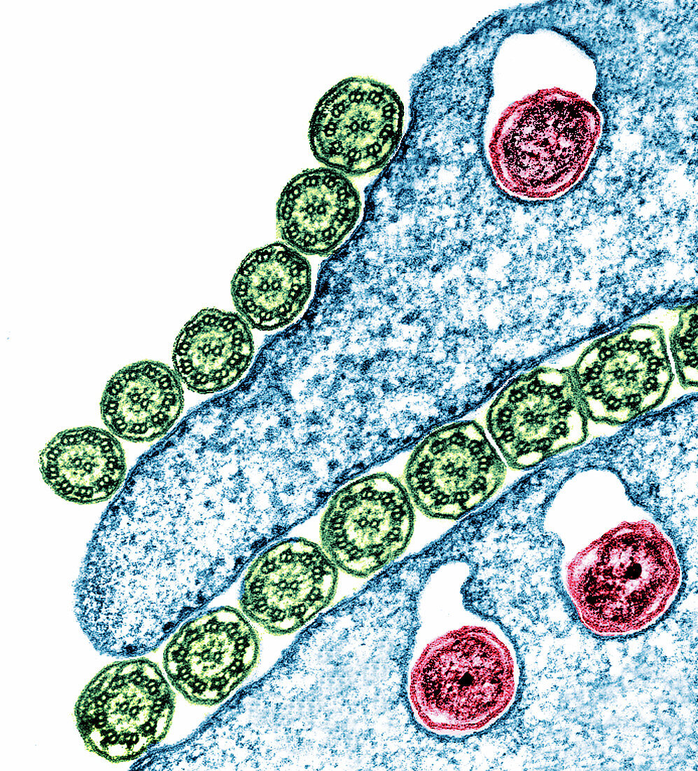
Trichonympha protozoan section,TEM
Bildnummer 11905650

| Trichonympha protozoan. Coloured transmission electron micrograph (TEM) of a section through part of a Trichonympha sp. flagellate protozoan. This protozoan inhabits the guts of termites,where it is responsible for digesting cellulose in the wood that they eat. Flagellate protozoans bear numerous flagella,or cilia,hair-like structures that they beat for motility. Several of these are seen sectioned here (green). Each cilium contains 20 microtubules (black circles),arranged as a central pair surrounded by nine other pairs. The red bodies in the protozoan are symbiotic bacteria. Magnification: x40,000 when printed 10cm tall | |
| Lizenzart: | Lizenzpflichtig |
| Credit: | Science Photo Library |
| Bildgröße: | 2411 px × 2661 px |
| Modell-Rechte: | nicht erforderlich |
| Eigentums-Rechte: | nicht erforderlich |
| Restrictions: | - |
Preise für dieses Bild ab 15 €
Universitäten & Organisationen
(Informationsmaterial Digital, Informationsmaterial Print, Lehrmaterial Digital etc.)
ab 15 €
Redaktionell
(Bücher, Bücher: Sach- und Fachliteratur, Digitale Medien (redaktionell) etc.)
ab 30 €
Werbung
(Anzeigen, Aussenwerbung, Digitale Medien, Fernsehwerbung, Karten, Werbemittel, Zeitschriften etc.)
ab 55 €
Handelsprodukte
(bedruckte Textilie, Kalender, Postkarte, Grußkarte, Verpackung etc.)
ab 75 €
Pauschalpreise
Rechtepakete für die unbeschränkte Bildnutzung in Print oder Online
ab 495 €
Keywords
- Axonem,
- Bakterien,
- Bakterium,
- Biologie,
- biologisch,
- Darm,
- farbig,
- Fauna,
- Flagellen,
- gefärbt,
- Geißel,
- Geißeltierchen,
- Mikrobiologie,
- mikrobiologisch,
- Mikrotubuli,
- Protozoen,
- Protozoon,
- Sektion,
- sektioniert,
- Symbiose,
- symbiotisch,
- tem,
- Termite,
- Tier,
- Tierwelt,
- Transmissionselektronenmikroskop,
- Wimpern,
- wirbellos,
- Wirbellose
