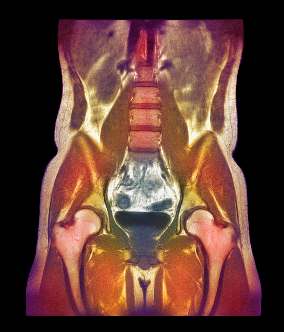
Abdominal scan,MRI
Bildnummer 11877107

| Abdominal scan. Coloured magnetic resonance imaging (MRI) scan of a woman's abdomen in coronal (frontal) section. The vertebrae (pale brown upper centre to centre) which form the spinal column are separated by the cartilaginous intervertebral discs (horizontal dark brown lines). Below the spine is the bladder (dark blue black,lower centre),which receives urine from the kidneys (one seen at upper left,brown oval) and stores it before it is excreted. On either side of the bladder lie the femurs (thigh bones,pink white,lower left and lower right,running to bottom). These are the longest and strongest bones in the body | |
| Lizenzart: | Lizenzpflichtig |
| Credit: | Science Photo Library / Fraser, Simon |
| Bildgröße: | 2400 px × 2800 px |
| Modell-Rechte: | nicht erforderlich |
| Eigentums-Rechte: | nicht erforderlich |
| Restrictions: | - |
Preise für dieses Bild ab 15 €
Universitäten & Organisationen
(Informationsmaterial Digital, Informationsmaterial Print, Lehrmaterial Digital etc.)
ab 15 €
Redaktionell
(Bücher, Bücher: Sach- und Fachliteratur, Digitale Medien (redaktionell) etc.)
ab 30 €
Werbung
(Anzeigen, Aussenwerbung, Digitale Medien, Fernsehwerbung, Karten, Werbemittel, Zeitschriften etc.)
ab 55 €
Handelsprodukte
(bedruckte Textilie, Kalender, Postkarte, Grußkarte, Verpackung etc.)
ab 75 €
Pauschalpreise
Rechtepakete für die unbeschränkte Bildnutzung in Print oder Online
ab 495 €
