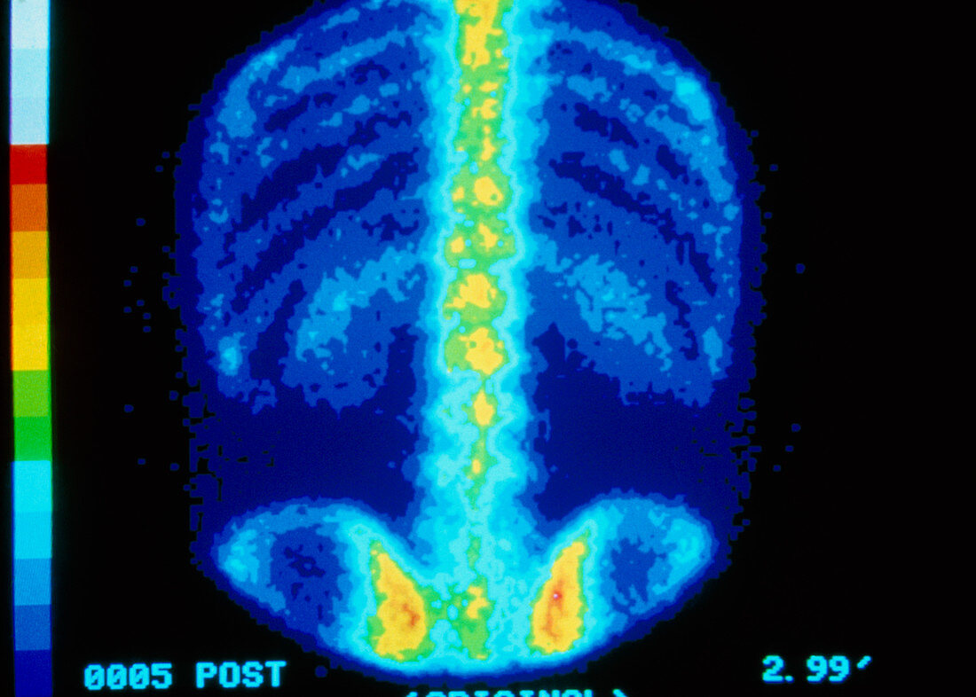
False-colour bone scintigram of a healthy adult
Bildnummer 11877054

| False colour bone scintigram (gamma camera scan) of a coronal (frontal) view of a normal adult skeleton,showing ribs,thoracic & lumbar spine and pelvis (bottom). Bone scans record the distribution & intensity of gamma radiations emanating from a radionuclide injected into the body prior to taking the scan,which concentrates in bone. A crystal scintillator in the camera resolves radiations as flashes of light that are detected by a system of photomultipliers. Bone scans are used to assess presence & extent of bone cancers,which present as sites of increased radionuclide uptake that appear as brighter,"hot spots" on the image | |
| Lizenzart: | Lizenzpflichtig |
| Credit: | Science Photo Library / CNRI |
| Bildgröße: | 3873 px × 2769 px |
| Modell-Rechte: | nicht erforderlich |
| Eigentums-Rechte: | nicht erforderlich |
| Restrictions: | - |
Preise für dieses Bild ab 15 €
Universitäten & Organisationen
(Informationsmaterial Digital, Informationsmaterial Print, Lehrmaterial Digital etc.)
ab 15 €
Redaktionell
(Bücher, Bücher: Sach- und Fachliteratur, Digitale Medien (redaktionell) etc.)
ab 30 €
Werbung
(Anzeigen, Aussenwerbung, Digitale Medien, Fernsehwerbung, Karten, Werbemittel, Zeitschriften etc.)
ab 55 €
Handelsprodukte
(bedruckte Textilie, Kalender, Postkarte, Grußkarte, Verpackung etc.)
ab 75 €
Pauschalpreise
Rechtepakete für die unbeschränkte Bildnutzung in Print oder Online
ab 495 €
