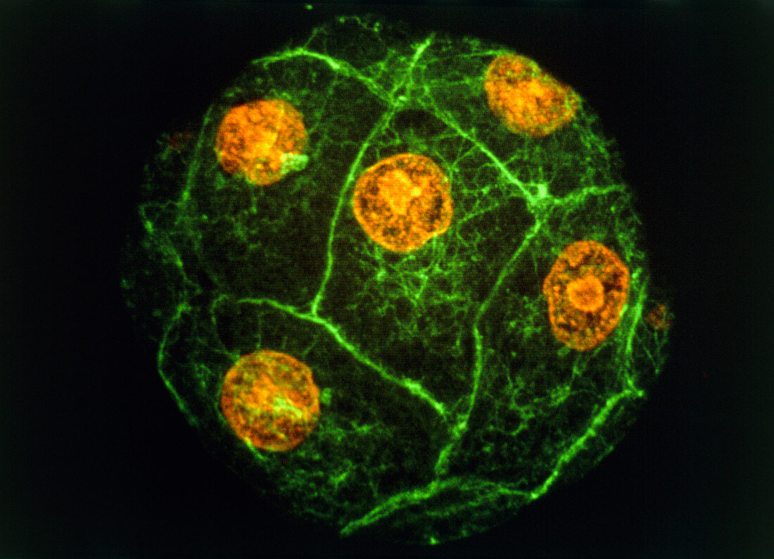
Sea urchin embryo
Bildnummer 11875505

| Sea urchin embryo. Immunofluorescence micrograph of a sea urchin embryo at the 8-16 cell stage. The orange structures are the cell nuclei. The bound- aries between different cells show up as green lines. Following fertilization an embryo undergoes multiple rounds cell division,dividing each time into double the number of cells (1,2,4,8 ...). Eventually a hollow ball of hundreds of cells is produced,which then starts to differentiate. This picture was made by exposing the embryo to fluor- escent antibodies that bind to certain proteins in the cell. The embryo was then viewed with a laser- scanning light microscope,which makes the anti- bodies fluoresce. Magnification unknown | |
| Lizenzart: | Lizenzpflichtig |
| Credit: | Science Photo Library / PROF. G. SCHATTEN |
| Bildgröße: | 5181 px × 3741 px |
| Modell-Rechte: | nicht erforderlich |
| Eigentums-Rechte: | nicht erforderlich |
| Restrictions: | - |
Preise für dieses Bild ab 15 €
Universitäten & Organisationen
(Informationsmaterial Digital, Informationsmaterial Print, Lehrmaterial Digital etc.)
ab 15 €
Redaktionell
(Bücher, Bücher: Sach- und Fachliteratur, Digitale Medien (redaktionell) etc.)
ab 30 €
Werbung
(Anzeigen, Aussenwerbung, Digitale Medien, Fernsehwerbung, Karten, Werbemittel, Zeitschriften etc.)
ab 55 €
Handelsprodukte
(bedruckte Textilie, Kalender, Postkarte, Grußkarte, Verpackung etc.)
ab 75 €
Pauschalpreise
Rechtepakete für die unbeschränkte Bildnutzung in Print oder Online
ab 495 €
