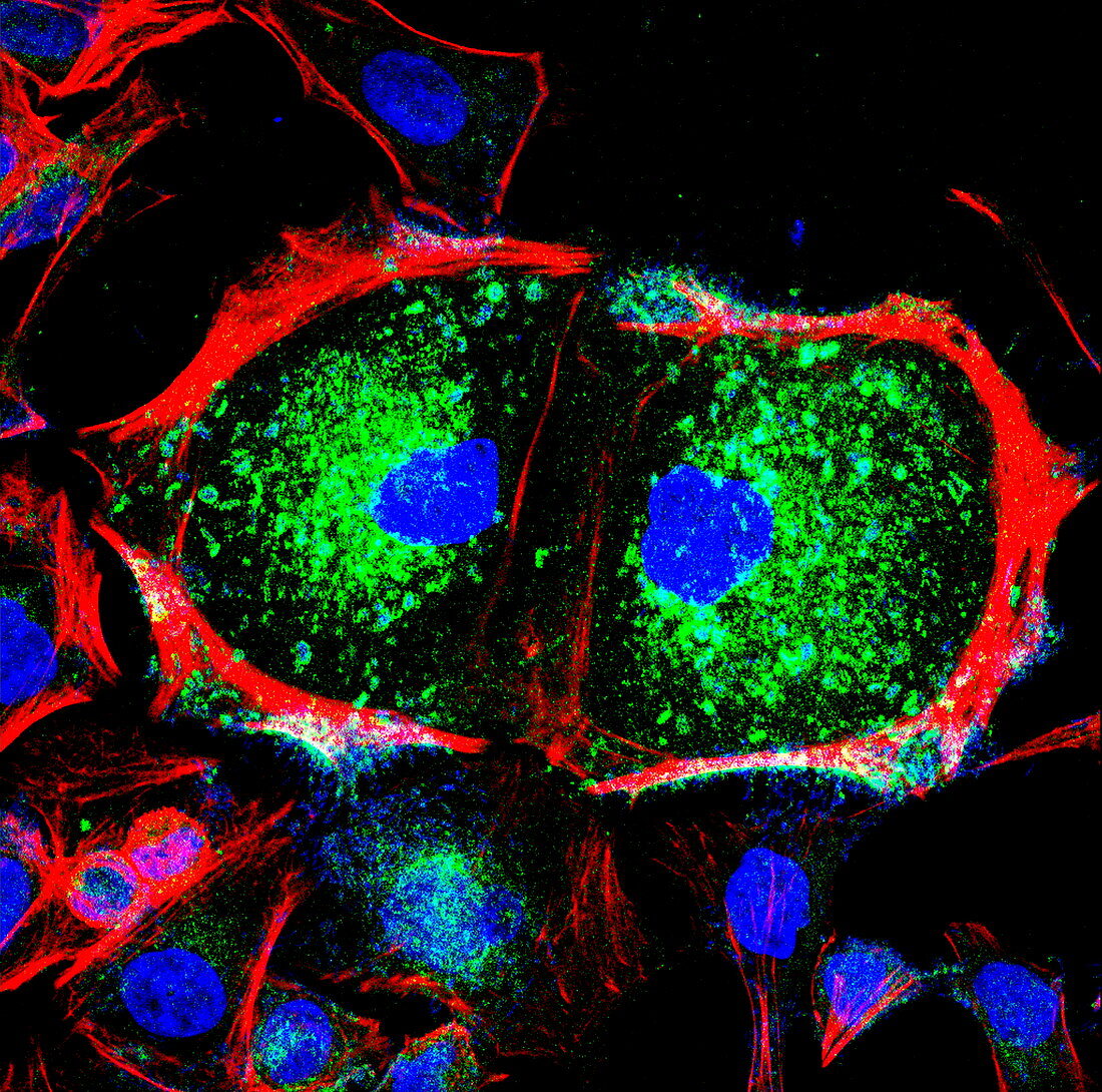
Dividing cell,light micrograph
Bildnummer 11874756

| Cell division. Fluorescent light micrograph of a cell that has divided by mitosis,the asexual replication of a cell into two new cells. This is late telophase,the mitosis stage where the cell's genetic material has separated and the cleavage plane (centre) has formed between the two new cells (left and right). The genetic material has been replicated and divided between two new cell nuclei (blue). The actin cytoskeleton (red) surrounds each cell. A receptor protein is marked in green. These are epithelial cells,cells that make up the body's internal and external surface layers,such as the skin and the walls of the intestines | |
| Lizenzart: | Lizenzpflichtig |
| Credit: | Science Photo Library |
| Bildgröße: | 2971 px × 2944 px |
| Modell-Rechte: | nicht erforderlich |
| Eigentums-Rechte: | nicht erforderlich |
| Restrictions: | - |
Preise für dieses Bild ab 15 €
Universitäten & Organisationen
(Informationsmaterial Digital, Informationsmaterial Print, Lehrmaterial Digital etc.)
ab 15 €
Redaktionell
(Bücher, Bücher: Sach- und Fachliteratur, Digitale Medien (redaktionell) etc.)
ab 30 €
Werbung
(Anzeigen, Aussenwerbung, Digitale Medien, Fernsehwerbung, Karten, Werbemittel, Zeitschriften etc.)
ab 55 €
Handelsprodukte
(bedruckte Textilie, Kalender, Postkarte, Grußkarte, Verpackung etc.)
ab 75 €
Pauschalpreise
Rechtepakete für die unbeschränkte Bildnutzung in Print oder Online
ab 495 €
Keywords
- Aktin,
- Atomkern,
- Biologie,
- biologisch,
- Desoxiribonukleinsäure,
- DNA,
- Duo,
- Einteilung,
- epithelial,
- Fluoreszenz,
- fluoreszierend,
- gesund,
- Histologie,
- histologisch,
- Kerne,
- kopierend,
- Lichtmikroskop,
- lichtmikroskopische Aufnahme,
- Mitose,
- normal,
- Paar,
- Replikation,
- Teilen,
- Zelle,
- Zellen,
- zellular,
- Zwei,
- Zytologie,
- Zytologisch,
- Zytoskelett
