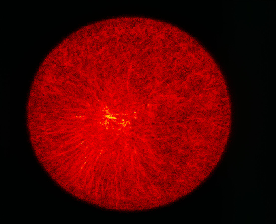
Confocal LM of a fertilised sea urchin egg
Bildnummer 11874468

| Fertilised egg (first of 3 images). Confocal light micrograph of a sea urchin egg after fertilisation showing migration of the pronuclei. Microtubules are red,radiating from a region (yellow) near the cell centre. These microtubules are helping to move the male and female pronuclei,the separate genetic material of sperm and egg (not clearly seen) towards the cell centre so that it can fuse. After it fuses into a "true" nucleus,the cell is termed a zygote and it can begin to divide to form an embryo. In confocal microscopy a fluorescent dye in the specimen is excited by laser light. See photo's P649/036 and P649/037 in this sequence. Magnification: x450 at 6x7cm size | |
| Lizenzart: | Lizenzpflichtig |
| Credit: | Science Photo Library / Whitaker, Michael |
| Bildgröße: | 4996 px × 4063 px |
| Modell-Rechte: | nicht erforderlich |
| Eigentums-Rechte: | nicht erforderlich |
| Restrictions: | - |
Preise für dieses Bild ab 15 €
Universitäten & Organisationen
(Informationsmaterial Digital, Informationsmaterial Print, Lehrmaterial Digital etc.)
ab 15 €
Redaktionell
(Bücher, Bücher: Sach- und Fachliteratur, Digitale Medien (redaktionell) etc.)
ab 30 €
Werbung
(Anzeigen, Aussenwerbung, Digitale Medien, Fernsehwerbung, Karten, Werbemittel, Zeitschriften etc.)
ab 55 €
Handelsprodukte
(bedruckte Textilie, Kalender, Postkarte, Grußkarte, Verpackung etc.)
ab 75 €
Pauschalpreise
Rechtepakete für die unbeschränkte Bildnutzung in Print oder Online
ab 495 €
