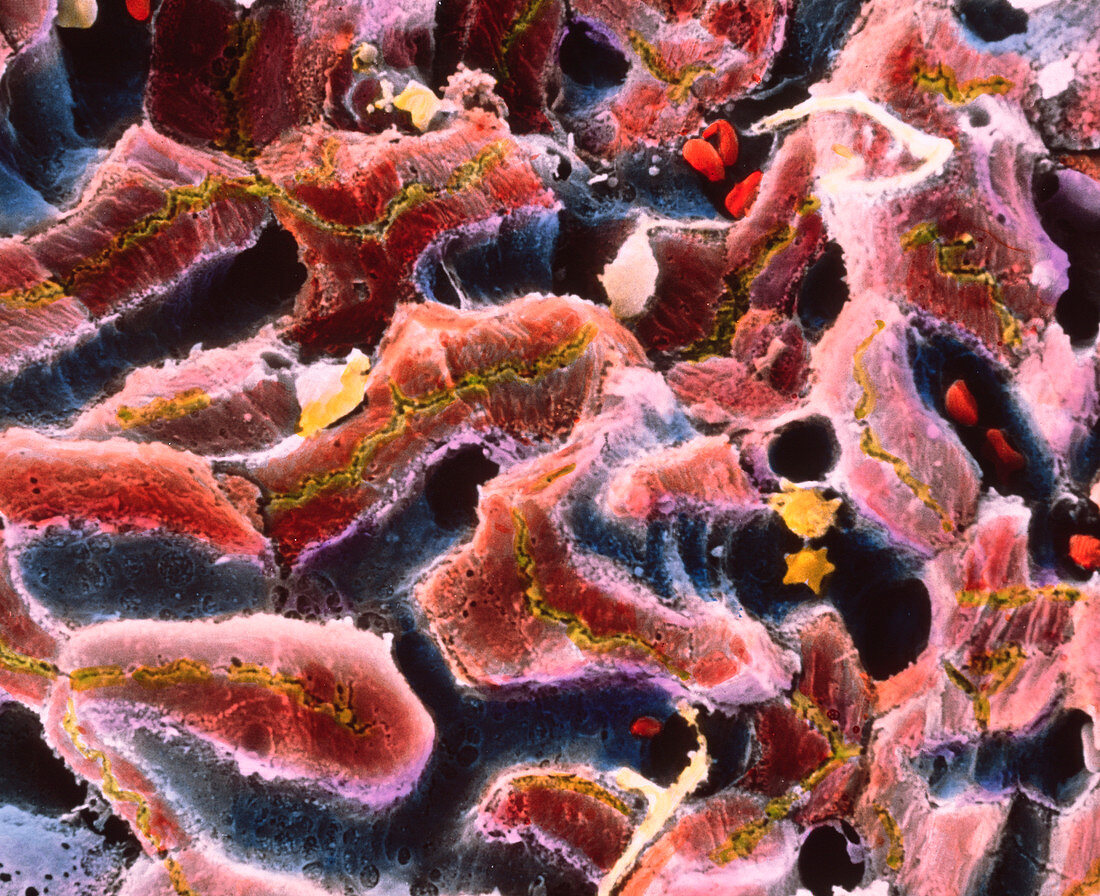
SEM of liver lobule
Bildnummer 11872873

| False-colour scanning electron micrograph (SEM) of the cell structure within a lobule of the liver. Hepatic cells (shades of brown) surround sinusoids (channels,blue) in a three-dimensional network. Blood cells (red) flow through these sinusoids to link up to a central vein. In this process of blood flow,liver cells are specialized to clean the blood of microorganisms,toxins,aged cells,and debris. Bile formed by liver cells is released into tiny bile capillaries (cholangioles,green). Bile then enters the gall bladder to be used for digestive purposes. The liver also synthesizes Vitamin A. Magnification: x1,050 at 6x7cm size. Magnification: x1,675 at 4x5 inch size | |
| Lizenzart: | Lizenzpflichtig |
| Credit: | Science Photo Library / UNIVERSITY LA SAPIENZA, ROME / DEPT. OF ANATOMY / PROF. P. MOTTA |
| Bildgröße: | 4157 px × 3385 px |
| Modell-Rechte: | nicht erforderlich |
| Eigentums-Rechte: | nicht erforderlich |
| Restrictions: | - |
Preise für dieses Bild ab 15 €
Universitäten & Organisationen
(Informationsmaterial Digital, Informationsmaterial Print, Lehrmaterial Digital etc.)
ab 15 €
Redaktionell
(Bücher, Bücher: Sach- und Fachliteratur, Digitale Medien (redaktionell) etc.)
ab 30 €
Werbung
(Anzeigen, Aussenwerbung, Digitale Medien, Fernsehwerbung, Karten, Werbemittel, Zeitschriften etc.)
ab 55 €
Handelsprodukte
(bedruckte Textilie, Kalender, Postkarte, Grußkarte, Verpackung etc.)
ab 75 €
Pauschalpreise
Rechtepakete für die unbeschränkte Bildnutzung in Print oder Online
ab 495 €
