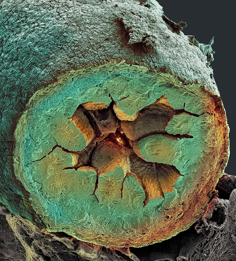
Small intestine,SEM
Bildnummer 11872755

| Small intestine. Coloured scanning electron micrograph (SEM) of a cross-section through a foetal small intestine. The small intestine runs from the stomach to the large intestine. It is where digestion is completed and nutrients and water are absorbed into the blood. The interior (lumen) of the small intestine is lined with a highly folded surface. The folds,known as villi,project into the lumen increasing the surface area for absorption. Beneath the villi is the submucosal layer,which contains blood vessels. Beyond the submucosa is a layer of smooth muscle,which contracts and relaxes to move food along the intestine. Magnification: x45 when printed 10 centimetres wide | |
| Lizenzart: | Lizenzpflichtig |
| Credit: | Science Photo Library / Gschmeissner, Steve |
| Bildgröße: | 3170 px × 3500 px |
| Modell-Rechte: | nicht erforderlich |
| Eigentums-Rechte: | nicht erforderlich |
| Restrictions: | - |
Preise für dieses Bild ab 15 €
Universitäten & Organisationen
(Informationsmaterial Digital, Informationsmaterial Print, Lehrmaterial Digital etc.)
ab 15 €
Redaktionell
(Bücher, Bücher: Sach- und Fachliteratur, Digitale Medien (redaktionell) etc.)
ab 30 €
Werbung
(Anzeigen, Aussenwerbung, Digitale Medien, Fernsehwerbung, Karten, Werbemittel, Zeitschriften etc.)
ab 55 €
Handelsprodukte
(bedruckte Textilie, Kalender, Postkarte, Grußkarte, Verpackung etc.)
ab 75 €
Pauschalpreise
Rechtepakete für die unbeschränkte Bildnutzung in Print oder Online
ab 495 €
Keywords
- Anatomie,
- Biologie,
- biologisch,
- Darm,
- Darm-,
- Dünndarm,
- eingefärbt,
- Falten,
- farbig,
- fötal,
- Gefaltet,
- gefärbt,
- Gewebe,
- Histologie,
- histologisch,
- Lumen,
- menschlicher Körper,
- Muskel,
- Querschnitt,
- Rasterelektronenmikroskop,
- rasterelektronenmikroskopische Aufnahme,
- REM,
- sektioniert,
- Submukosa,
- System,
- Trakt,
- vaskulär,
- Verdauung,
- Verdauungs-,
- Zotte,
- Zotten
