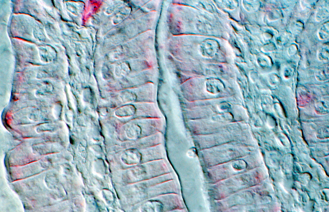
Intestinal cells,light micrograph
Bildnummer 11872749

| Intestinal cells. Light micrograph of a section through two folds (villi) in the small intestine. The villi are aligned vertically,with one at left and one at right. Each fold consists of an inner layer of supportive connective tissue (irregular structure),overlaid by an outer layer of columnar epithelial cells). The round structures are the nuclei of these cells. The folded nature of the villi increases the surface area for absorption of nutrients by the microvilli (not clearly seen) on the outer surface of the epithelial cells | |
| Lizenzart: | Lizenzpflichtig |
| Credit: | Science Photo Library |
| Bildgröße: | 3728 px × 2404 px |
| Modell-Rechte: | nicht erforderlich |
| Eigentums-Rechte: | nicht erforderlich |
| Restrictions: | - |
Preise für dieses Bild ab 15 €
Universitäten & Organisationen
(Informationsmaterial Digital, Informationsmaterial Print, Lehrmaterial Digital etc.)
ab 15 €
Redaktionell
(Bücher, Bücher: Sach- und Fachliteratur, Digitale Medien (redaktionell) etc.)
ab 30 €
Werbung
(Anzeigen, Aussenwerbung, Digitale Medien, Fernsehwerbung, Karten, Werbemittel, Zeitschriften etc.)
ab 55 €
Handelsprodukte
(bedruckte Textilie, Kalender, Postkarte, Grußkarte, Verpackung etc.)
ab 75 €
Pauschalpreise
Rechtepakete für die unbeschränkte Bildnutzung in Print oder Online
ab 495 €
Keywords
- Atomkern,
- Biologie,
- biologisch,
- Darm,
- Darm-,
- Dünndarm,
- Epithel,
- epithelial,
- Falten,
- Gedärme,
- gesund,
- Histologie,
- histologisch,
- Kerne,
- Lichtmikroskop,
- lichtmikroskopische Aufnahme,
- menschlicher Körper,
- normal,
- Oberfläche,
- Sektion,
- sektioniert,
- Verdauungs-,
- Zelle,
- Zellen,
- zellular,
- Zotte,
- Zotten,
- Zytologie,
- Zytologisch
