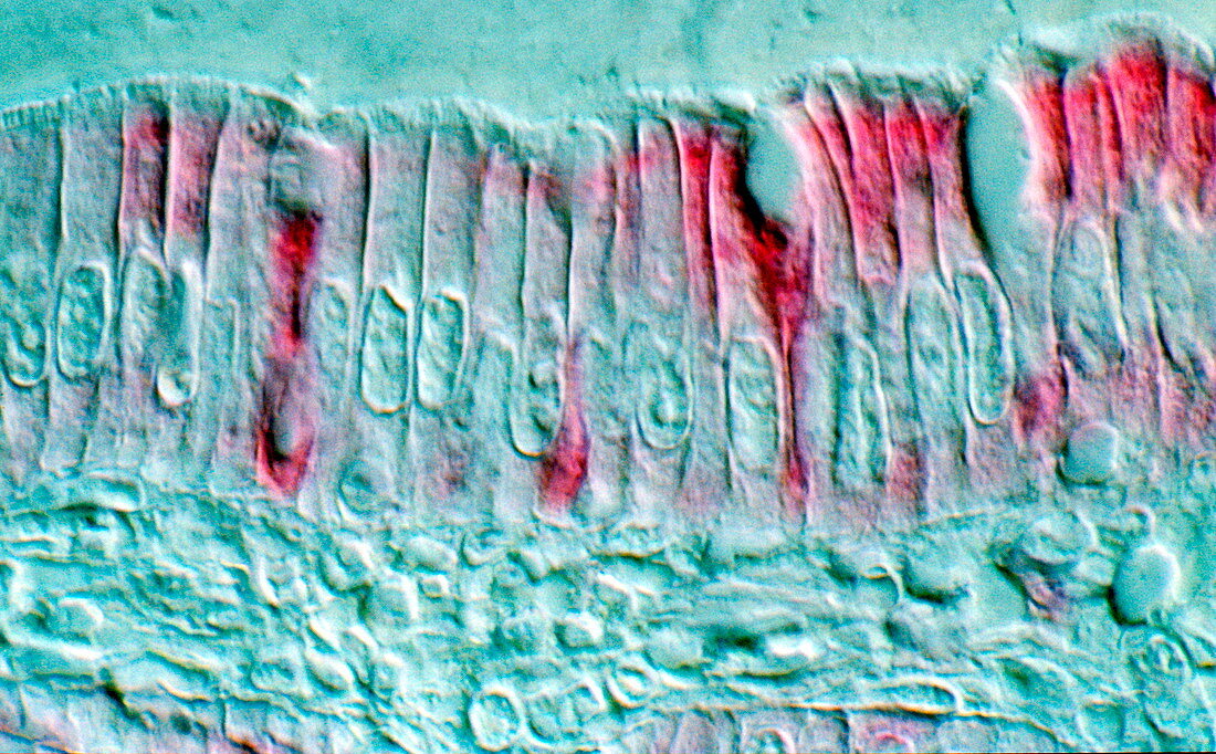
Intestinal cells,light micrograph
Bildnummer 11872748

| Intestinal cells. Light micrograph of a section through the wall of the small intestine. The upper layer is seen here,consisting of many columnar cells (columnar epithelial cells). The cell nuclei (oval) are seen about two-thirds of the way down the cells. On the outer surface of the cells (across top) is a brush-layer of many small hair- like absorptive structures called microvilli. These are exposed to food absorbing nutrients from it as it passes through the small intestine. Below the epithelial cells is a supporting layer of connective tissue (across bottom) | |
| Lizenzart: | Lizenzpflichtig |
| Credit: | Science Photo Library |
| Bildgröße: | 3404 px × 2112 px |
| Modell-Rechte: | nicht erforderlich |
| Eigentums-Rechte: | nicht erforderlich |
| Restrictions: | - |
Preise für dieses Bild ab 15 €
Universitäten & Organisationen
(Informationsmaterial Digital, Informationsmaterial Print, Lehrmaterial Digital etc.)
ab 15 €
Redaktionell
(Bücher, Bücher: Sach- und Fachliteratur, Digitale Medien (redaktionell) etc.)
ab 30 €
Werbung
(Anzeigen, Aussenwerbung, Digitale Medien, Fernsehwerbung, Karten, Werbemittel, Zeitschriften etc.)
ab 55 €
Handelsprodukte
(bedruckte Textilie, Kalender, Postkarte, Grußkarte, Verpackung etc.)
ab 75 €
Pauschalpreise
Rechtepakete für die unbeschränkte Bildnutzung in Print oder Online
ab 495 €
Keywords
- Atomkern,
- Bilder,
- Biologie,
- biologisch,
- Darm,
- Darm-,
- Dünndarm,
- Epithel,
- epithelial,
- Fächer,
- Gedärme,
- gesund,
- Histologie,
- histologisch,
- Kerne,
- Lichtmikroskop,
- lichtmikroskopische Aufnahme,
- menschlicher Körper,
- mikroskopische Fotos,
- Mikrovilli,
- normal,
- Oberfläche,
- Sektion,
- sektioniert,
- Verdauungs-,
- vergrößertes Bild,
- Zelle,
- Zellen,
- zellular,
- Zytologie,
- Zytologisch
