False-colour SEM of epithelium in the oesophagus
Bildnummer 11872558
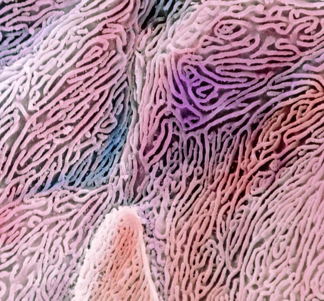
| False-colour scanning electron micrograph (SEM) of the epithelium in the oesophagus. Individual epithelial cells are seen here,each with a highly folded surface. These microfolds are called micro- plicae. At lower centre left,an obvious boundary to one cell can be seen. This stratified squamous epithelium consists of flattened cells that occur many layers thick. As a muscular non-absorptive tube,the oesophagus transports swallowed food to the stomach. The microfolds on the cells keep the oesophagus moist; trap mucous to lubricate passing food; and strengthen the epithelium against food abrasion. Magnification: x2,900 at 6x7cm size. Magnification: x4,650 at 4x5 inch size | |
| Lizenzart: | Lizenzpflichtig |
| Credit: | Science Photo Library / UNIVERSITY LA SAPIENZA, ROME / DEPT. OF ANATOMY / PROF. P. MOTTA |
| Bildgröße: | 4382 px × 4073 px |
| Modell-Rechte: | nicht erforderlich |
| Eigentums-Rechte: | nicht erforderlich |
| Restrictions: | - |
Preise für dieses Bild ab 15 €
Universitäten & Organisationen
(Informationsmaterial Digital, Informationsmaterial Print, Lehrmaterial Digital etc.)
ab 15 €
Redaktionell
(Bücher, Bücher: Sach- und Fachliteratur, Digitale Medien (redaktionell) etc.)
ab 30 €
Werbung
(Anzeigen, Aussenwerbung, Digitale Medien, Fernsehwerbung, Karten, Werbemittel, Zeitschriften etc.)
ab 55 €
Handelsprodukte
(bedruckte Textilie, Kalender, Postkarte, Grußkarte, Verpackung etc.)
ab 75 €
Pauschalpreise
Rechtepakete für die unbeschränkte Bildnutzung in Print oder Online
ab 495 €