Purkinje nerve cells,light micrograph
Bildnummer 11871423
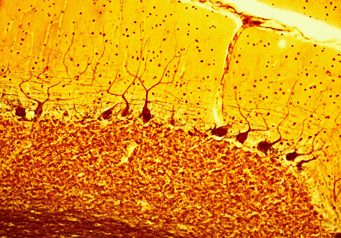
| Purkinje nerve cells. Light micrograph of a section through the cerebellum in the brain,showing Purkinje nerve cells. Each Purkinje cell is composed of a flask-shaped cell body (brown dots) from which branch numerous dendrites (brown strands). Purkinje cells are arranged at the junction of the molecular (yellow) and granular (brown) layers of the cerebellum,which make up the grey matter of the brain. Nerve impulses flow to the Purkinje cells through their dendrites. The message is then passed on to the white matter deep in the cerebellar cortex. Nuclei (small dots) of other nerve cells,such as glial cells,can be seen throughout the grey matter. Magnification: x110 when 10cm wide | |
| Lizenzart: | Lizenzpflichtig |
| Credit: | Science Photo Library / Gschmeissner, Steve |
| Bildgröße: | 4224 px × 2950 px |
| Modell-Rechte: | nicht erforderlich |
| Eigentums-Rechte: | nicht erforderlich |
| Restrictions: | - |
Preise für dieses Bild ab 15 €
Universitäten & Organisationen
(Informationsmaterial Digital, Informationsmaterial Print, Lehrmaterial Digital etc.)
ab 15 €
Redaktionell
(Bücher, Bücher: Sach- und Fachliteratur, Digitale Medien (redaktionell) etc.)
ab 30 €
Werbung
(Anzeigen, Aussenwerbung, Digitale Medien, Fernsehwerbung, Karten, Werbemittel, Zeitschriften etc.)
ab 55 €
Handelsprodukte
(bedruckte Textilie, Kalender, Postkarte, Grußkarte, Verpackung etc.)
ab 75 €
Pauschalpreise
Rechtepakete für die unbeschränkte Bildnutzung in Print oder Online
ab 495 €
Keywords
- Anatomie,
- anatomisch,
- Atomkern,
- Biologie,
- biologisch,
- Braun,
- Dendrit,
- diagonal,
- Gehirn,
- Gelb,
- Grau,
- Histologie,
- histologisch,
- Kerne,
- Kleinhirn,
- Lichtmikroskop,
- lichtmikroskopische Aufnahme,
- Mensch,
- Menschen,
- menschlicher Körper,
- molekulare Schicht,
- Nervensystem,
- Nervenzelle,
- neural,
- Neurologie,
- neurologisch,
- Neuron,
- neuronale,
- Neuronen,
- Person,
- Querschnitt,
- Scheibe,
- Sektion,
- weiße Substanz,
- Zelle,
- Zellen,
- zentrales Nervensystem