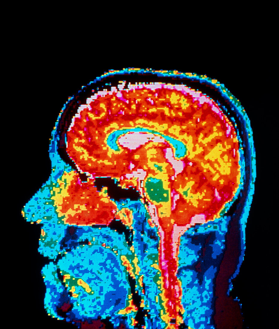
NMR image showing structures of the brain
Bildnummer 11870690

| False-colour nuclear magnetic resonance (NMR) image of a sagittal section through the human head,showing structures of a normal brain. The cerebrum (the 2 cerebral hemispheres) which forms the bulk of brain tissue appears orange-red. The pink areas at the centre,top,and surrounding the spinal cord at bottom represent cerebro-spinal fluid (CSF); the central pink area shows CSF in the ventricles (chambers) at the centre of the brain. The curved blue area surrounding the ventricles is the corpus callosum,a structure connecting the 2 cerebral hemispheres. At bottom right (top right of spinal cord) is the cerebell- um,the centre of balance & muscular coordination | |
| Lizenzart: | Lizenzpflichtig |
| Credit: | Science Photo Library |
| Bildgröße: | 4200 px × 4961 px |
| Modell-Rechte: | nicht erforderlich |
| Eigentums-Rechte: | nicht erforderlich |
| Restrictions: | - |
Preise für dieses Bild ab 15 €
Universitäten & Organisationen
(Informationsmaterial Digital, Informationsmaterial Print, Lehrmaterial Digital etc.)
ab 15 €
Redaktionell
(Bücher, Bücher: Sach- und Fachliteratur, Digitale Medien (redaktionell) etc.)
ab 30 €
Werbung
(Anzeigen, Aussenwerbung, Digitale Medien, Fernsehwerbung, Karten, Werbemittel, Zeitschriften etc.)
ab 55 €
Handelsprodukte
(bedruckte Textilie, Kalender, Postkarte, Grußkarte, Verpackung etc.)
ab 75 €
Pauschalpreise
Rechtepakete für die unbeschränkte Bildnutzung in Print oder Online
ab 495 €
