Heartbeat,MRI
Bildnummer 11869168

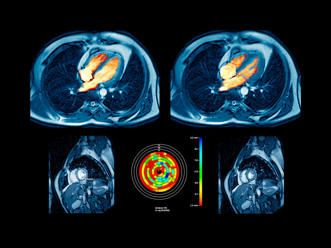
| Heartbeat. Coloured magnetic resonance imaging (MRI) scans of transverse (top) and vertical (bottom) sections through a 45-year-old's chest showing a healthy heart. The two ventricles (lower chambers,orange) of the heart are shown. The width of the left ventricle has been measured. At left the heart is in ventricular diastole,a period of relaxation. As the muscles relax the pressure in the left ventricle decreases and oxygenated blood enters it from the left atrium (upper chamber). At right the heart is in systole. The left ventricle is contracting,pushing the blood into the aorta,the body's main artery | |
| Lizenzart: | Lizenzpflichtig |
| Credit: | Science Photo Library / Zephyr |
| Bildgröße: | 4535 px × 3401 px |
| Modell-Rechte: | nicht erforderlich |
| Eigentums-Rechte: | nicht erforderlich |
| Restrictions: | - |
Preise für dieses Bild ab 15 €
Universitäten & Organisationen
(Informationsmaterial Digital, Informationsmaterial Print, Lehrmaterial Digital etc.)
ab 15 €
Redaktionell
(Bücher, Bücher: Sach- und Fachliteratur, Digitale Medien (redaktionell) etc.)
ab 30 €
Werbung
(Anzeigen, Aussenwerbung, Digitale Medien, Fernsehwerbung, Karten, Werbemittel, Zeitschriften etc.)
ab 55 €
Handelsprodukte
(bedruckte Textilie, Kalender, Postkarte, Grußkarte, Verpackung etc.)
ab 75 €
Pauschalpreise
Rechtepakete für die unbeschränkte Bildnutzung in Print oder Online
ab 495 €
Keywords
- 40er Jahre,
- Anatomie,
- anatomisch,
- diagonal,
- Entspannung,
- Erwachsene,
- farbig,
- gefärbt,
- gesund,
- Herz,
- Herzen,
- Herzkreislaufsystem,
- Herzschlag,
- Kammer,
- Kontraktion,
- Lungen,
- Magnetresonanztomografie,
- menschlicher Körper,
- Messung,
- MRT-Untersuchung,
- Muskel,
- normal,
- Organ,
- Scanner,
- Sektion,
- sektioniert,
- Thorax,
- Truhe,
- Ventrikel,
- Vierziger Jahre
