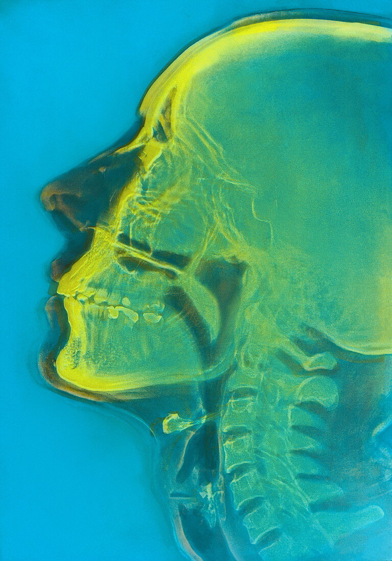
Human skull,X-ray
Bildnummer 11868036

| False-colour X-ray image of a human skull,showing a profile view featuring the jaw & upper spinal region. The image was obtained using xerography,a technique where a xerox plate is used to record transmitted X-rays instead of photographic film; soft tissues of the nose & around the throat are visible. Also prominent are the airways from the nose & mouth passing into the trachea or windpipe; these appear as dark grey passages. The cervical vertebrae - the spinal bones in the neck - are seen; the bony processes on their dorsal surface (left) have a saw-toothed appearance | |
| Lizenzart: | Lizenzpflichtig |
| Credit: | Science Photo Library / CNRI |
| Bildgröße: | 3193 px × 4576 px |
| Modell-Rechte: | nicht erforderlich |
| Eigentums-Rechte: | nicht erforderlich |
| Restrictions: | - |
Preise für dieses Bild ab 15 €
Universitäten & Organisationen
(Informationsmaterial Digital, Informationsmaterial Print, Lehrmaterial Digital etc.)
ab 15 €
Redaktionell
(Bücher, Bücher: Sach- und Fachliteratur, Digitale Medien (redaktionell) etc.)
ab 30 €
Werbung
(Anzeigen, Aussenwerbung, Digitale Medien, Fernsehwerbung, Karten, Werbemittel, Zeitschriften etc.)
ab 55 €
Handelsprodukte
(bedruckte Textilie, Kalender, Postkarte, Grußkarte, Verpackung etc.)
ab 75 €
Pauschalpreise
Rechtepakete für die unbeschränkte Bildnutzung in Print oder Online
ab 495 €
