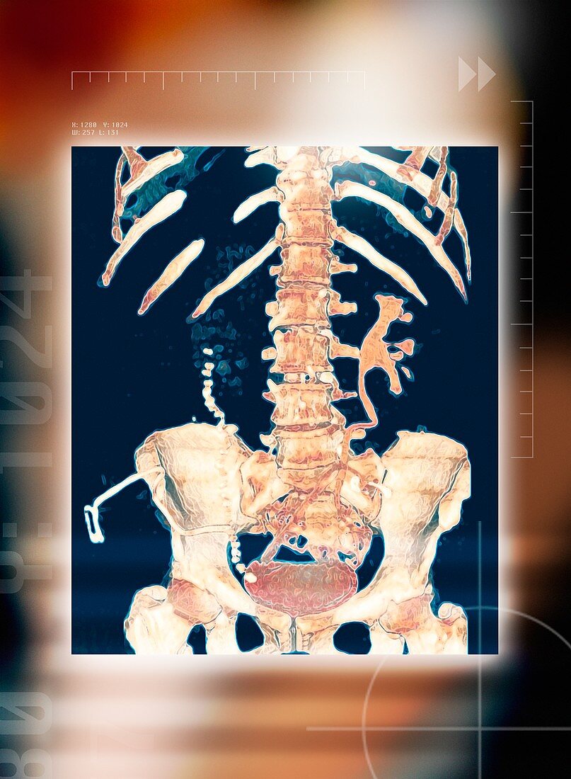
Abdominal bones,3-D CT scan
Bildnummer 11867878

| Abdominal bones,coloured 3-D computer tomography (CT) scan. The lower part of the ribcage is in upper frame,with the pelvis in lower frame. They are connected by the spine,which runs down centre. The spine is made up of block-shaped bones called vertebrae. At the base of the pelvis is the bladder (red oval). The bladder is full of urine,which is seen as the patient was given an intravenous contrast medium to highlight structures on an X-ray. The contrast medium is excreted in urine. One kidney is seen at centre right (branched structure). Urine drains from the kidney to the bladder through a ureter. CT scans use X-rays to build up an image of body structures | |
| Lizenzart: | Lizenzpflichtig |
| Credit: | Science Photo Library / Maslo, Miriam |
| Bildgröße: | 3580 px × 4882 px |
| Modell-Rechte: | nicht erforderlich |
| Eigentums-Rechte: | nicht erforderlich |
| Restrictions: | - |
Preise für dieses Bild ab 15 €
Universitäten & Organisationen
(Informationsmaterial Digital, Informationsmaterial Print, Lehrmaterial Digital etc.)
ab 15 €
Redaktionell
(Bücher, Bücher: Sach- und Fachliteratur, Digitale Medien (redaktionell) etc.)
ab 30 €
Werbung
(Anzeigen, Aussenwerbung, Digitale Medien, Fernsehwerbung, Karten, Werbemittel, Zeitschriften etc.)
ab 55 €
Handelsprodukte
(bedruckte Textilie, Kalender, Postkarte, Grußkarte, Verpackung etc.)
ab 75 €
Pauschalpreise
Rechtepakete für die unbeschränkte Bildnutzung in Print oder Online
ab 495 €
Keywords
- 3-d,
- Abdomen,
- Anatomie,
- anatomisch,
- Ausscheidung,
- Bauch,
- Becken,
- Blase,
- Brustkorb,
- Computertomographie,
- CT-Scan,
- Dreidimensional,
- farbig,
- gefärbt,
- gesund,
- Harnleiter,
- Knochen,
- Kontrastmittel,
- Lendenwirbelsäule,
- Medizin,
- medizinisch,
- menschlicher Körper,
- Niere,
- Nieren-,
- normal,
- Organ,
- pelvin,
- Physiologie,
- physiologisch,
- Rippe,
- Rippen,
- Röntgen,
- Röntgengerät,
- Scanner,
- Skelett,
- Skelett-,
- System,
- unterer Rücken,
- Voll,
- Wirbel,
- Wirbelsäule,
- Wirbelsäulen-
