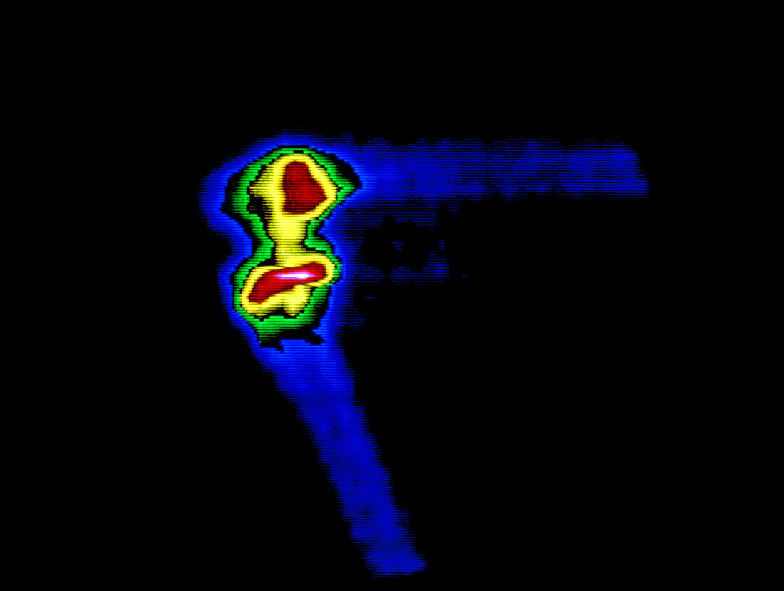
F/colour gamma scan of a normal human knee joint
Bildnummer 11867629

| Healthy knee joint. False-colour computer enhanced gamma scan (scintigram) of a human knee joint seen in side view. At top (blue),the thigh femur bone is positioned horizontally and connects at the knee joint to the tibia shin bone (blue,lower centre). The articular surfaces of these bones meet in cartilage (red),and the joint is united by ligaments (yellow). This image shows the dis- tribution of a radioactive tracer technetium-99m,injected into the subject & which concentrates in bone. Radiation is recorded by a gamma camera. Bone scanning is frequently used in screening for secondary disease in cancer patients. Cancerous bone would appear as brighter "hot spots" | |
| Lizenzart: | Lizenzpflichtig |
| Credit: | Science Photo Library / CNRI |
| Bildgröße: | 5292 px × 3990 px |
| Modell-Rechte: | nicht erforderlich |
| Eigentums-Rechte: | nicht erforderlich |
| Restrictions: | - |
Preise für dieses Bild ab 15 €
Universitäten & Organisationen
(Informationsmaterial Digital, Informationsmaterial Print, Lehrmaterial Digital etc.)
ab 15 €
Redaktionell
(Bücher, Bücher: Sach- und Fachliteratur, Digitale Medien (redaktionell) etc.)
ab 30 €
Werbung
(Anzeigen, Aussenwerbung, Digitale Medien, Fernsehwerbung, Karten, Werbemittel, Zeitschriften etc.)
ab 55 €
Handelsprodukte
(bedruckte Textilie, Kalender, Postkarte, Grußkarte, Verpackung etc.)
ab 75 €
Pauschalpreise
Rechtepakete für die unbeschränkte Bildnutzung in Print oder Online
ab 495 €
