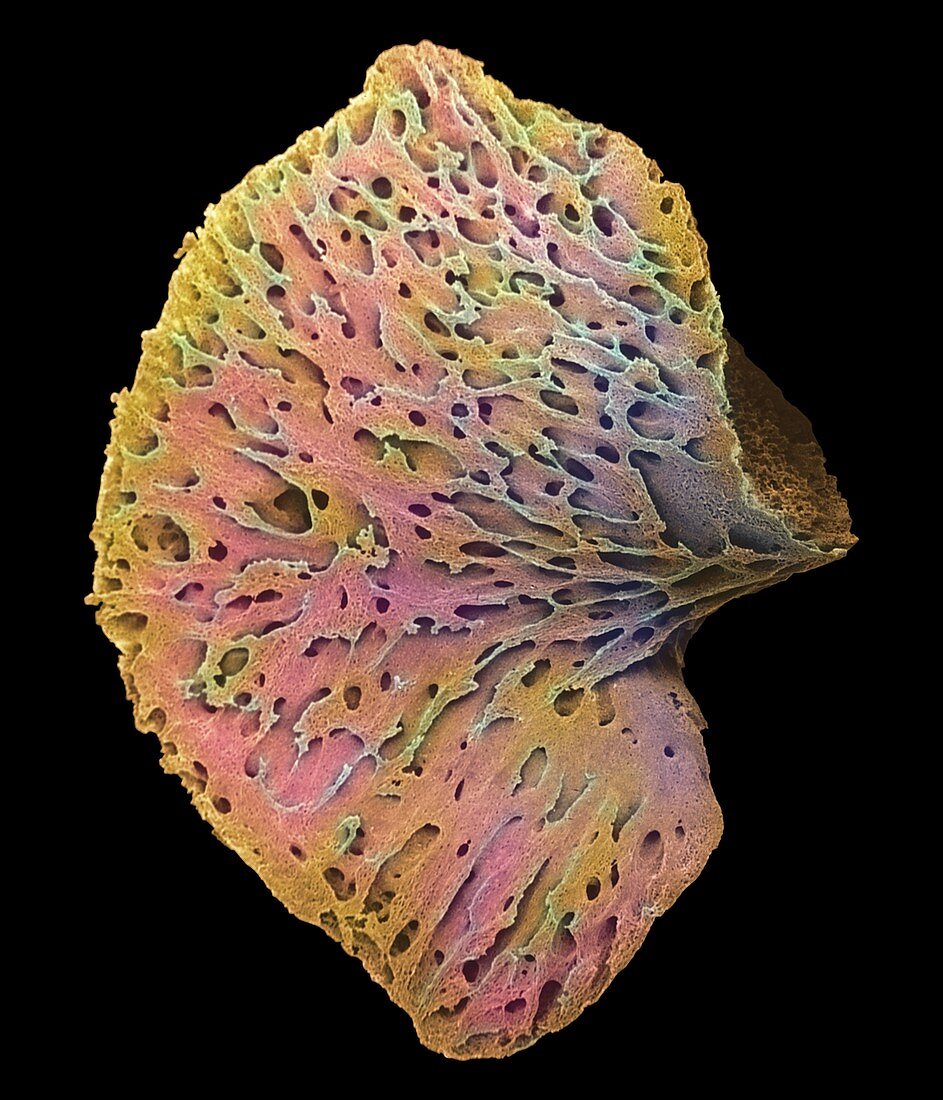
Foetal bone
Bildnummer 11867467

| Foetal bone. Coloured scanning electron micrograph (SEM) of a pelvic bone of an 11 week old foetus. Only the calcified matrix of the bone remains as the cartilage has been lost during the processing of the sample. The pelvis is initially formed of cartilage in the foetus,but it is steadily replaced by bone as the foetus grows. Some cartilage is still present in childrens' bones. Magnification: x12.5 at 6x7cm size | |
| Lizenzart: | Lizenzpflichtig |
| Credit: | Science Photo Library / Moscoso, Dr. G. |
| Bildgröße: | 2400 px × 2800 px |
| Modell-Rechte: | nicht erforderlich |
| Eigentums-Rechte: | nicht erforderlich |
| Restrictions: | - |
Preise für dieses Bild ab 15 €
Universitäten & Organisationen
(Informationsmaterial Digital, Informationsmaterial Print, Lehrmaterial Digital etc.)
ab 15 €
Redaktionell
(Bücher, Bücher: Sach- und Fachliteratur, Digitale Medien (redaktionell) etc.)
ab 30 €
Werbung
(Anzeigen, Aussenwerbung, Digitale Medien, Fernsehwerbung, Karten, Werbemittel, Zeitschriften etc.)
ab 55 €
Handelsprodukte
(bedruckte Textilie, Kalender, Postkarte, Grußkarte, Verpackung etc.)
ab 75 €
Pauschalpreise
Rechtepakete für die unbeschränkte Bildnutzung in Print oder Online
ab 495 €
