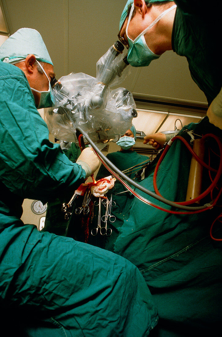
Split beam: operating microscope used in surgery
Bildnummer 11853228

| Surgeons using a "split beam" operating microscope to remove a brain tumour. The microscope is wrapped in plastic to maintain sterile conditions,& allows the surgeons a view of the same illuminated field: it is also fitted with a video camera so that students may follow the course of the operation. The surgeon at left is using an ultrasound probe to liquify and remove the tumour. Ultrasonic vibrations emulsify the growth,allowing it to be sucked away. Furthermore,tissue is removed from its centre,causing the growth to collapse in on itself,and allowing any remaining tumour matter to be more easily teased away from surrounding healthy tissue | |
| Lizenzart: | Lizenzpflichtig |
| Credit: | Science Photo Library / Dohrn, Martin |
| Bildgröße: | 3365 px × 5109 px |
| Modell-Rechte: | Derzeit liegt noch kein Release vor. Bitte kontaktieren Sie uns vor Verwendung. |
| Eigentums-Rechte: | nicht erforderlich |
| Restrictions: | - |
Preise für dieses Bild ab 15 €
Universitäten & Organisationen
(Informationsmaterial Digital, Informationsmaterial Print, Lehrmaterial Digital etc.)
ab 15 €
Redaktionell
(Bücher, Bücher: Sach- und Fachliteratur, Digitale Medien (redaktionell) etc.)
ab 30 €
Werbung
(Anzeigen, Aussenwerbung, Digitale Medien, Fernsehwerbung, Karten, Werbemittel, Zeitschriften etc.)
ab 55 €
Handelsprodukte
(bedruckte Textilie, Kalender, Postkarte, Grußkarte, Verpackung etc.)
ab 75 €
Pauschalpreise
Rechtepakete für die unbeschränkte Bildnutzung in Print oder Online
ab 495 €
