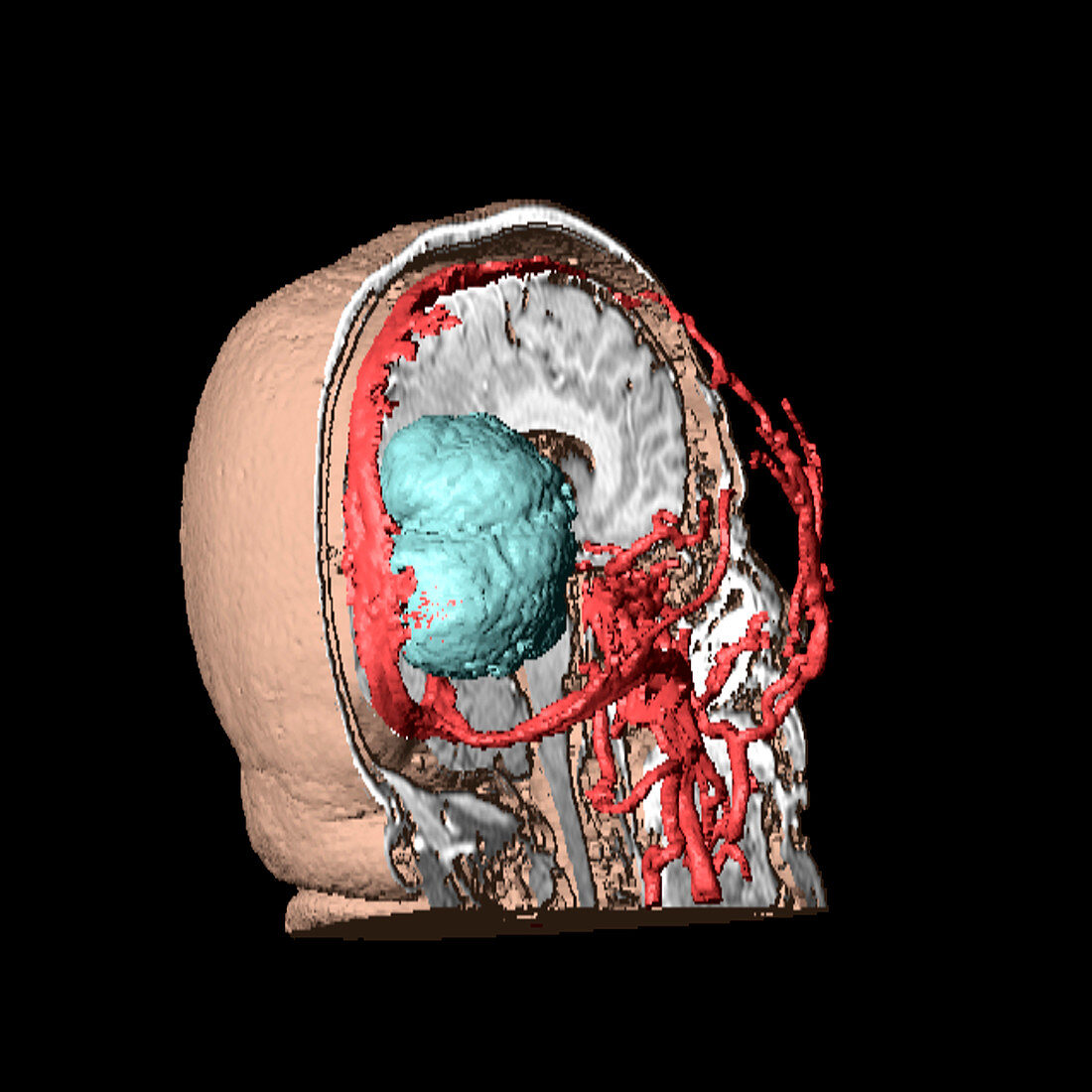
Brain tumour,3D scan
Bildnummer 11841169

| Brain tumour. 3D model of the head (rear view) of a 59-year-old woman,made up from numerous MRI (magnetic resonance imaging) scans. It is showing the internal structures of the right-hand side of the head,revealing the brain (white),and blood vessels (red). A large brain tumour,a benign (non-cancerous) meningioma,can be seen in blue at the back of the brain. Meningiomas are most often benign and arise from the meninges,the protective membranes that cover the brain and spinal cord. As it grows,a meningioma compresses adjacent brain tissue,and symptoms,such as headaches,are often related to this compression. Usually the tumour can be removed surgically,although radiotherapy may also be needed | |
| Lizenzart: | Lizenzpflichtig |
| Credit: | Science Photo Library / Fraser, Simon |
| Bildgröße: | 2965 px × 2965 px |
| Modell-Rechte: | nicht erforderlich |
| Eigentums-Rechte: | nicht erforderlich |
| Restrictions: | - |
Preise für dieses Bild ab 15 €
Universitäten & Organisationen
(Informationsmaterial Digital, Informationsmaterial Print, Lehrmaterial Digital etc.)
ab 15 €
Redaktionell
(Bücher, Bücher: Sach- und Fachliteratur, Digitale Medien (redaktionell) etc.)
ab 30 €
Werbung
(Anzeigen, Aussenwerbung, Digitale Medien, Fernsehwerbung, Karten, Werbemittel, Zeitschriften etc.)
ab 55 €
Handelsprodukte
(bedruckte Textilie, Kalender, Postkarte, Grußkarte, Verpackung etc.)
ab 75 €
Pauschalpreise
Rechtepakete für die unbeschränkte Bildnutzung in Print oder Online
ab 495 €
Keywords
- 3 dimensional,
- 3-d,
- 3-dimensional,
- 3D,
- 50er Jahre,
- Biologie,
- biologisch,
- Blutgefäß,
- Cutaway,
- Diagnose,
- Dreidimensional,
- Erwachsene,
- Frau,
- Fünfziger Jahre,
- geduldig,
- Gefäße,
- Gehirn,
- gutartig,
- Hirnhaut,
- Kopf,
- Krankheit,
- Magnetresonanztomografie,
- Medizin,
- medizinisch,
- Meningiom,
- Mensch,
- menschlicher Körper,
- mittleren Alters,
- Modell-,
- MRT-Untersuchung,
- Neuroimaging,
- Neurologie,
- neurologisch,
- Onkologie,
- onkologisch,
- Rückansicht,
- Scan,
- Scanner,
- Schädel,
- Scheibe,
- Sektion,
- Tumor,
- Wachstum,
- Weiblich
