Spine disorder
Bildnummer 11840891

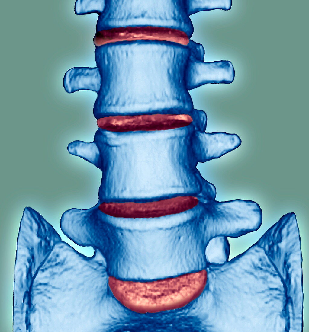
| Spine disorder. Frontal coloured 3D magnetic resonance imaging (MRI) scan of the lumbar (lower) spine of a 28 year old patient with lordosis,an exaggerated inward curvature of the spine. The blue blocks are the vertebrae,the orange areas between them are the intervertebral spaces. The sacrum is at the bottom. The last intervertebral space (bottom) is abnormally large. This indicates lordosis. If the curvature is flexible,it corrects itself when the patient bends forwards,no treatment is needed and the condition will not progress or cause problems. If the curvature is fixed and it does not correct itself,surgery may be needed | |
| Lizenzart: | Lizenzpflichtig |
| Credit: | Science Photo Library / Zephyr |
| Bildgröße: | 3402 px × 3658 px |
| Modell-Rechte: | nicht erforderlich |
| Eigentums-Rechte: | nicht erforderlich |
| Restrictions: | - |
Preise für dieses Bild ab 15 €
Universitäten & Organisationen
(Informationsmaterial Digital, Informationsmaterial Print, Lehrmaterial Digital etc.)
ab 15 €
Redaktionell
(Bücher, Bücher: Sach- und Fachliteratur, Digitale Medien (redaktionell) etc.)
ab 30 €
Werbung
(Anzeigen, Aussenwerbung, Digitale Medien, Fernsehwerbung, Karten, Werbemittel, Zeitschriften etc.)
ab 55 €
Handelsprodukte
(bedruckte Textilie, Kalender, Postkarte, Grußkarte, Verpackung etc.)
ab 75 €
Pauschalpreise
Rechtepakete für die unbeschränkte Bildnutzung in Print oder Online
ab 495 €
Keywords
- 20er Jahre,
- 3-d,
- 3D,
- abnormal,
- Computertomographie,
- CT-Scan,
- Dreidimensional,
- eingefärbt,
- farbig,
- Frontal,
- Gebogen,
- gefärbt,
- Kondition,
- Krankheit,
- Kreuzbein,
- Krümmung,
- Kurve,
- Lendenwirbelsäule,
- Medizin,
- medizinisch,
- Mensch,
- Raum,
- Räume,
- Rücken,
- Rückgrat,
- Scanner,
- Scheibe,
- Störung,
- Wirbel,
- Wirbelsäule,
- Wirbelsäulen-,
- zwanziger Jahre
