Heart disease
Bildnummer 11839932

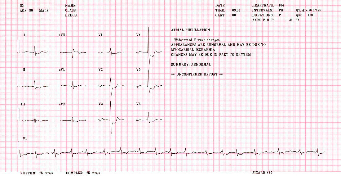
| Heart disease. Electrocardiogram (ECG) showing the heartbeat during atrial fibrillation in an 80- year-old man. An ECG records the pumping of the heart's chambers,tracing electrical waves (brown) that are the output from 12 external electrodes (I-III,V1-V6 and aVR,aVL and aVF). In atrial fibrillation,the heart muscle fibres of the atria (two of the heart chambers) fail to contract in a co-ordinated way,leading to a rapid,irregular beat. This case may have been caused by myocardial ischaemia,inadequate blood flow to the heart muscle due to blocked blood vessels. Abnormalities in this ECG include changes (e.g. trough in trace V4) in the T wave after the main heartbeat peak | |
| Lizenzart: | Lizenzpflichtig |
| Credit: | Science Photo Library / Marazzi, Dr. P. |
| Bildgröße: | 3500 px × 1805 px |
| Modell-Rechte: | nicht erforderlich |
| Eigentums-Rechte: | nicht erforderlich |
| Restrictions: | - |
Preise für dieses Bild ab 15 €
Universitäten & Organisationen
(Informationsmaterial Digital, Informationsmaterial Print, Lehrmaterial Digital etc.)
ab 15 €
Redaktionell
(Bücher, Bücher: Sach- und Fachliteratur, Digitale Medien (redaktionell) etc.)
ab 30 €
Werbung
(Anzeigen, Aussenwerbung, Digitale Medien, Fernsehwerbung, Karten, Werbemittel, Zeitschriften etc.)
ab 55 €
Handelsprodukte
(bedruckte Textilie, Kalender, Postkarte, Grußkarte, Verpackung etc.)
ab 75 €
Pauschalpreise
Rechtepakete für die unbeschränkte Bildnutzung in Print oder Online
ab 495 €
