Heart disease
Bildnummer 11839919

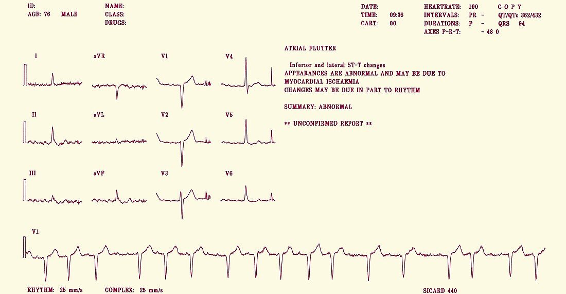
| Heart disease. Electrocardiogram (ECG) of the heartbeat of a 76-year-old man with an atrial flutter and myocardial ischaemia. An ECG measures the pumping of the heart chambers using 12 external electrodes (I-III,V1-V6 and aVR,aVL and aVF),recording it as electrical waves (purple). A flutter is an increased rate of contraction of the upper heart chambers (atria),seen as numerous shallow waves (for example in traces I and II). It may be caused by myocardial ischaemia,inadequate blood flow to the heart caused by narrowed vessels. This is revealed by the S-T wave depressions (pronounced troughs after tall peaks) in the traces for electrodes II,III and aVF | |
| Lizenzart: | Lizenzpflichtig |
| Credit: | Science Photo Library / Marazzi, Dr. P. |
| Bildgröße: | 3500 px × 1816 px |
| Modell-Rechte: | nicht erforderlich |
| Eigentums-Rechte: | nicht erforderlich |
| Restrictions: | - |
Preise für dieses Bild ab 15 €
Universitäten & Organisationen
(Informationsmaterial Digital, Informationsmaterial Print, Lehrmaterial Digital etc.)
ab 15 €
Redaktionell
(Bücher, Bücher: Sach- und Fachliteratur, Digitale Medien (redaktionell) etc.)
ab 30 €
Werbung
(Anzeigen, Aussenwerbung, Digitale Medien, Fernsehwerbung, Karten, Werbemittel, Zeitschriften etc.)
ab 55 €
Handelsprodukte
(bedruckte Textilie, Kalender, Postkarte, Grußkarte, Verpackung etc.)
ab 75 €
Pauschalpreise
Rechtepakete für die unbeschränkte Bildnutzung in Print oder Online
ab 495 €
