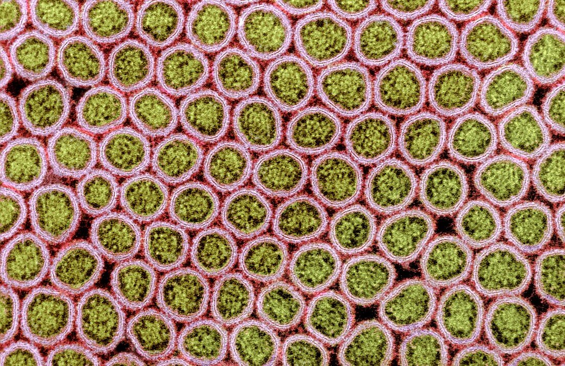
Intestinal microvilli,SEM
Bildnummer 11634976

| Intestinal microvilli. Coloured transmission electron micrograph (SEM) of a transverse section through microvilli,showing their interiors. These tiny hair-like structures form a dense brush-like covering on the absorptive surfaces of the cells lining the small intestine. The core of each microvillus is formed of filaments (green dots) with a glycolyx of polysaccharides surrounding the cell membrane (pink ring). Magnification: x50,000 when printed at 10 centimetres wide | |
| Lizenzart: | Lizenzpflichtig |
| Credit: | Science Photo Library / Gschmeissner, Steve |
| Bildgröße: | 3709 px × 2403 px |
| Modell-Rechte: | nicht erforderlich |
| Restrictions: | - |
Preise für dieses Bild ab 15 €
Universitäten & Organisationen
(Informationsmaterial Digital, Informationsmaterial Print, Lehrmaterial Digital etc.)
ab 15 €
Redaktionell
(Bücher, Bücher: Sach- und Fachliteratur, Digitale Medien (redaktionell) etc.)
ab 30 €
Werbung
(Anzeigen, Aussenwerbung, Digitale Medien, Fernsehwerbung, Karten, Werbemittel, Zeitschriften etc.)
ab 55 €
Handelsprodukte
(bedruckte Textilie, Kalender, Postkarte, Grußkarte, Verpackung etc.)
ab 75 €
Pauschalpreise
Rechtepakete für die unbeschränkte Bildnutzung in Print oder Online
ab 495 €
Keywords
- Absorption,
- Anatomie,
- anatomisch,
- Biologie,
- biologisch,
- Darm,
- Darm-,
- Dünndarm,
- eingefärbt,
- Ernährung,
- Farbig,
- Futter,
- Gedärme,
- gefärbt,
- gesund,
- GI tract,
- Innere,
- Kern,
- Magen-Darm-System,
- mehrere,
- Membran,
- menschlicher Körper,
- Mikrovilli,
- Mikrovillus,
- Nährstoffe,
- normal,
- REM,
- sektioniert,
- Struktur,
- Trakt,
- Transmissionselektronenmikroskop,
- transmissionselektronenmikroskopische Aufnahme,
- Transversalschnitt,
- Verdauung,
- Verdauungssystem,
- viele,
- Zellbilogie,
- Zellen,
- Zytologie,
- Zytologisch
