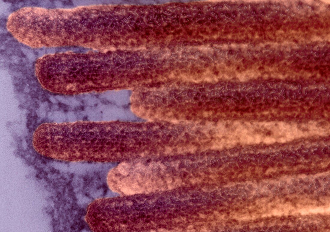
Intestinal microvilli,TEM
Bildnummer 11623770

| Intestinal microvilli. Transmission electron micrograph (TEM) of a section through an epithelial cell from a human jejunum (part of the small intestine),showing the densely packed microvilli. Each microvillus is approximately 1 micrometre long by 0.1 micrometres in diameter and contains a core of actin microfilaments. These tiny structures form a dense brush-like covering on the absorptive surfaces of the cells lining the small intestine. The cells absorb nutrients from digested food through the microvilli | |
| Lizenzart: | Lizenzpflichtig |
| Credit: | Science Photo Library / AMI Images |
| Bildgröße: | 3517 px × 2481 px |
| Modell-Rechte: | nicht erforderlich |
| Restrictions: | - |
Preise für dieses Bild ab 15 €
Universitäten & Organisationen
(Informationsmaterial Digital, Informationsmaterial Print, Lehrmaterial Digital etc.)
ab 15 €
Redaktionell
(Bücher, Bücher: Sach- und Fachliteratur, Digitale Medien (redaktionell) etc.)
ab 30 €
Werbung
(Anzeigen, Aussenwerbung, Digitale Medien, Fernsehwerbung, Karten, Werbemittel, Zeitschriften etc.)
ab 55 €
Handelsprodukte
(bedruckte Textilie, Kalender, Postkarte, Grußkarte, Verpackung etc.)
ab 75 €
Pauschalpreise
Rechtepakete für die unbeschränkte Bildnutzung in Print oder Online
ab 495 €
Keywords
- Absorption,
- Anatomie,
- anatomisch,
- Biologie,
- biologisch,
- Darm,
- Darm-,
- Dünndarm,
- eingefärbt,
- Ernährung,
- Futter,
- Gedärme,
- gefärbt,
- gesund,
- GI tract,
- Jejunum,
- Magen-Darm-System,
- mehrere,
- menschlicher Körper,
- Mikrovilli,
- Mikrovillus,
- Nährstoffe,
- normal,
- REM,
- Sektion,
- sektioniert,
- Trakt,
- Transmissionselektronenmikroskop,
- transmissionselektronenmikroskopische Aufnahme,
- Verdauung,
- Verdauungssystem,
- viele
