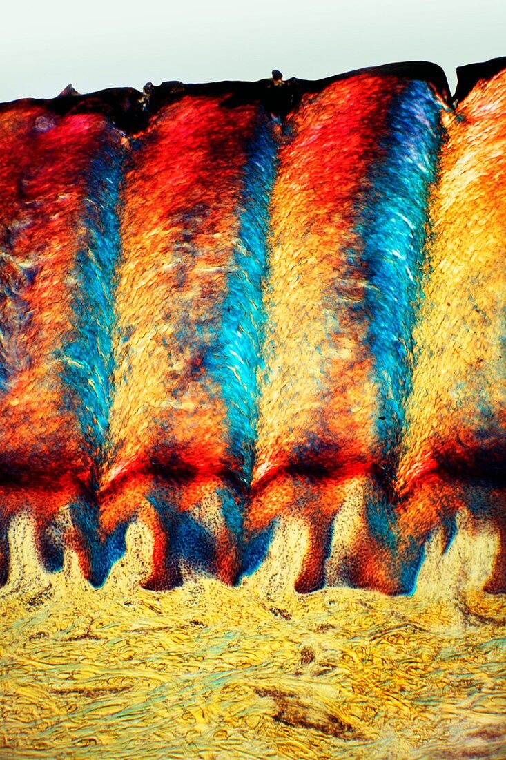
Heel skin tissue,light micrograph
Bildnummer 11549639

| Heel skin tissue. Polarised light micrograph of a transverse section through skin from the heel of a human foot. The sole of the foot has to withstand the weight of the body and friction when the foot pushes against the substratum. To cope with this,the outer epidermis forms deep layers of dead keratinised cells (red and blue) which are replaced from the meristematic Malpighian layer as they get worn away. The dermis (yellow and green) consists of collagenous elastic fibres. Magnification: x103 when printed at 10 centimetres high | |
| Lizenzart: | Lizenzpflichtig |
| Credit: | Science Photo Library / Wheeler, Dr. Keith |
| Bildgröße: | 3433 px × 5150 px |
| Modell-Rechte: | nicht erforderlich |
| Eigentums-Rechte: | nicht erforderlich |
| Restrictions: | - |
Preise für dieses Bild ab 15 €
Universitäten & Organisationen
(Informationsmaterial Digital, Informationsmaterial Print, Lehrmaterial Digital etc.)
ab 15 €
Redaktionell
(Bücher, Bücher: Sach- und Fachliteratur, Digitale Medien (redaktionell) etc.)
ab 30 €
Werbung
(Anzeigen, Aussenwerbung, Digitale Medien, Fernsehwerbung, Karten, Werbemittel, Zeitschriften etc.)
ab 55 €
Handelsprodukte
(bedruckte Textilie, Kalender, Postkarte, Grußkarte, Verpackung etc.)
ab 75 €
Pauschalpreise
Rechtepakete für die unbeschränkte Bildnutzung in Print oder Online
ab 495 €
Keywords
- Anatomie,
- anatomisch,
- Biologie,
- biologisch,
- dermal,
- Dermatologie,
- dermatologisch,
- Dick,
- epidermal,
- Epidermis,
- Fuß,
- gesund,
- Gewebe,
- Haut,
- Histologie,
- histologisch,
- Kollagen,
- Lichtmikroskop,
- lichtmikroskopische Aufnahme,
- Mensch,
- normal,
- Oberfläche,
- Person,
- polarisiert,
- Querschnitt,
- Sektion,
- sektioniert,
- Tief,
- Tot
