Protozoan (Vorticella sp.), SEM
Bildnummer 12377551
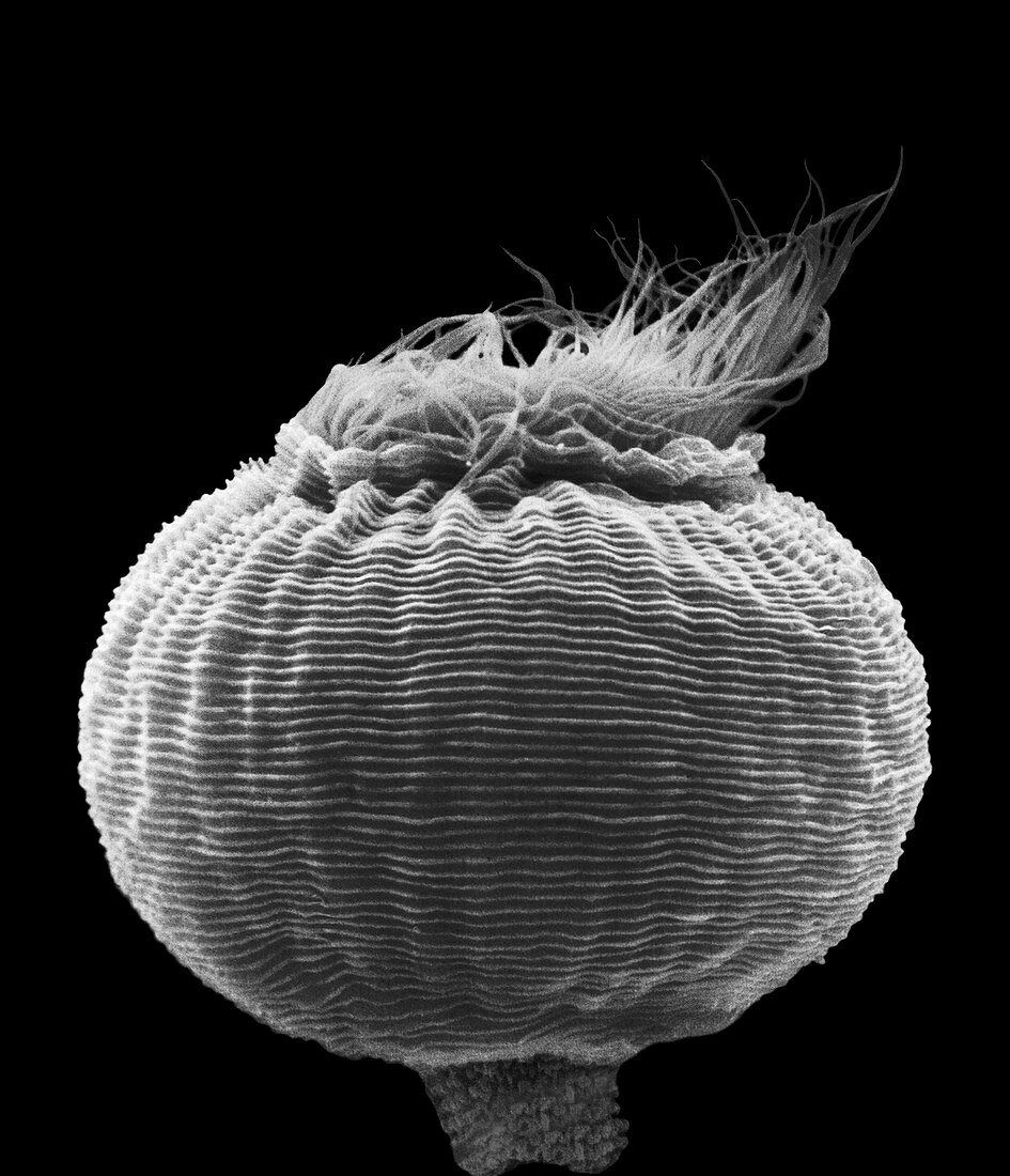
| Vorticella sp., ciliated protozoan, scanning electron micrograph (SEM). Shown is the main cell region of this stalked (attached) protozoan. The mouth is partially opened with cilia protruding. Vorticella species are stalked, inverted bell-shaped ciliates, placed among the peritrichs (peritrichous ciliates). Each cell has a separate stalk anchored onto the substrate, which contains a contractile fibril called a myoneme. When stimulated, this shortens, causing the stalk to coil like a spring. Vorticella species mainly live in freshwater ponds and streams. Magnification: x695 when shortest axis printed at 25 millimetres. | |
| Lizenzart: | Lizenzpflichtig |
| Credit: | Science Photo Library / DENNIS KUNKEL MICROSCOPY |
| Bildgröße: | 2943 px × 3427 px |
| Modell-Rechte: | nicht erforderlich |
| Eigentums-Rechte: | nicht erforderlich |
| Restrictions: | - |
Preise für dieses Bild ab 15 €
Universitäten & Organisationen
(Informationsmaterial Digital, Informationsmaterial Print, Lehrmaterial Digital etc.)
ab 15 €
Redaktionell
(Bücher, Bücher: Sach- und Fachliteratur, Digitale Medien (redaktionell) etc.)
ab 30 €
Werbung
(Anzeigen, Aussenwerbung, Digitale Medien, Fernsehwerbung, Karten, Werbemittel, Zeitschriften etc.)
ab 55 €
Handelsprodukte
(bedruckte Textilie, Kalender, Postkarte, Grußkarte, Verpackung etc.)
ab 75 €
Pauschalpreise
Rechtepakete für die unbeschränkte Bildnutzung in Print oder Online
ab 495 €
Keywords
- Ciliat,
- Ciliaten,
- Einfarbig,
- Elektron,
- Eukaryot,
- farbig,
- frisch,
- gefärbt,
- kontraktil,
- Marine,
- Meer,
- Mikrofotografie,
- Mund,
- Niemand,
- oligohymenophorea,
- Peritrich,
- Protozoen,
- Protozoon,
- rasterelektronenmikroskopische Aufnahme,
- REM,
- Salz,
- Scannen,
- schwarz und weiß,
- sessilida,
- Stengel,
- Süßwasser,
- Vorticella,
- Wasser,
- Wasser-,
- Wimpern